- Table View
- List View

Bone Tumours in Man and Animals
by L. N. OwenBone Tumours in Man and Animals covers advances in the understanding of the pathogenesis of bone neoplasia in humans and animals as well as their diagnosis and treatment. In the case of animals, particular emphasis is placed on dogs. This book is comprised of 15 chapters and begins by introducing the reader to the principles of diagnosis, including biopsy and bone scanning, and methods of treatment employed for bone tumors in human and animal patients, including surgery, radiotherapy, and chemotherapy. The following chapters focus on bone cysts and benign tumors of osseous origin; osteosarcoma in humans and animals such as dogs, cats, and mice; and tumors of cartilaginous origin such as solitary enchondroma, enchondromatosis, chondroblastoma, and chondromyxoid fibroma. Tumors arising from fibrous, fatty, neurological, or undifferentiated connective tissue are also considered, along with those arising from bone marrow or lymphoid tissue. This book concludes by offering suggestions for future research. This text is intended for clinicians, radiologists, and pathologists, both medical and veterinary, as well as for biologists, biochemists, and radiation physicists.

Bones: General Practice - The Integrative Approach Series
by Kerryn Phelps Craig HassedBones - General Practice: The Integrative Approach. Bones are complex organs with many important functions, the most obvious being structural. They provide support for the body and the means by which muscles can insert into fixed structures in order to allow movement. They are also important in hearing, through the transduction of sound via the ear’s ossicles, and they protect other soft organs that are easily damaged, such as the brain, eyes, kidneys, lungs and spleen. Bone marrow, which is largely within the medulla of the long bones, is the centre for production of blood cells (haematopoesis) and an important site for storage of fatty acids. Bones also have important metabolic functions. This chapter describes the following conditions affecting bones: osteoporosis, rickets, osteomalacia, Paget’s disease and bone cancer.

Bones: Orthopaedic Pathologies in Roman Imperial Age
by Andrea Piccioli Valentina Gazzaniga Paola CatalanoThis book presents the results of a unique macroscopic and radiological analysis, by X-ray and CT scan, of the bone pathologies of about 1800 subjects who lived at the time of the Roman Empire (first and second centuries A.D.) and whose remains were recovered during the excavation of a suburban necropolis of Rome. The survey, which represents a collaboration between the Italian Society of Orthopaedics and Traumatology and the Special Superintendent for the Archaeological Heritage of Rome, has yielded incredible images of different orthopaedic diseases in a period when no surgical treatment was available: there are cases of infection (osteomyelitis), metabolic disease (gout), hematologic disease (multiple myeloma), traumatic lesions and their complications and degenerative pathology (osteoarthritis, particularly secondary and overload). A multidisciplinary team including orthopaedists, paleopathologists, radiologists and medical historians has evaluated the major groups of bone disease in the population finding out incredible cases and picture of ortho-traumatologic pathologies in a pre-surgical era. The homogeneity of the sample and the number of subjects make this a study of fundamental importance.

Bones and Joints: 170 Radiological Exercises for Students and Practitioners (Exercises in Radiological Diagnosis)
by Michel RungeOsteoarticular pathology is a very frequent motive for consultation. Very often, the diagnosis relies upon symptomatology, and the physi cian requires confirmatory radiological investigations. Whatever the clinical indication, the interpretation of radiological data must be very rigorous. On the basis of a complete description of the radiographic images, according to a systematic analysis plan, a certain number of diagnostic hypotheses may be proposed. Selection of the most likely hypothesis requires the correlation of clinical, biological, and radiological data, and may sometimes necessi tate additional investigations, such as tomograms, scintigrams, and computed tomography (CT). 1 Part One Iconography 3 3 1 2 4 5 5 6 6 7 8 b a 8 a 9 10 11 12 10 13 14 11 15 a b 12 a c 13 17 b a c 14 15 c 16 17 23 21 a 22 b 18 19 20 21 22 23 33 34 24 25 37 38 26 27 40- 43 28 29 46 30 48 47 31 49 50 32 33 52 a b c 34 53 a b d c 35 54 a b 36 37 55 a 38 55 b c 39 56 57 40 58 41 60 61 42 43 63 64 44 65 66 45 67 68 46 69 a b 47 70 71 48 73 49 74 75 50 76 77 51 78 79 52 80 a b c 53 81 82 54 83 84 55 85 86 S6 87 88 57 89 90 58 91 92

Bones and Joints - E-book: A Guide for Students
by Chris GunnThis book is a clear, concise introduction to the subject which covers all the major bones and joints in the body in a logical and systematic way to aid understanding. The three generic chapters at the start of the book, covering an overview of bone, joints and pathology, provide the basic information required to ensure that the student is able to gain the most benefit from the subsequent area-specific chapters. The text is written in note form and the drawings are as clear and simple as possible so that they can be easily reproduced by students. In this edition a number of the radiographic images have been improved and replaced and the number of imaging techniques has increased by including PET and SPECT images New to this edition Improved clarity of the joint images A number of new radiographic images Insight Boxes Inclusion of PET and SPECT colour images Bones and Joints may be used as part of a self-directed learning programme by students examining and studying the real bones of the skeleton along with the images. It can also be used as part of a revision programme or as a reference text. It is aimed at all health care students who needs a good understanding of the skeletal system.

Bones and Joints - E-Book: A Guide for Students
by Chris GunnBones and Joints offers a clear and concise introduction to the bones and joints of the body along with pathology. Heavily illustrated with clear annotations, this is an essential learning, revision aid and reference for all radiography students and other health care students including nurses. Laid out in a logical and systemic way the text is easy to understand with brand new colour illustrations throughout. The three generic chapters at the start of the book, covering an overview of bone, joints and pathology, provide the basic information required to ensure that the student is able to gain the most benefit from the subsequent area-specific chapters. This is an essential book for all health care students who need a good understanding of the skeletal system. Now fully illustrated in colour throughout with clear annotations for easy understanding. Joint illustrations are colour coded to aid learning

Bones and Joints - E-Book: Bones and Joints - E-Book
by James HarcusNow in its eighth edition, this highly respected core textbook is essential reading for all healthcare students learning about the bones and joints of the body. The information is logically ordered and easy to read; comprehensive enough for students and health professionals alike, but not so dense as to be overwhelming. It covers the normal structure of bones and joints and goes on to provide an introduction to common fractures and pathology and how they appear on imaging. Bones and Joints is the perfect initial textbook, as well as a revision and refresher guide that will suit students of radiography, physiotherapy, osteology, sports medicine and nursing. - Clear and concise introduction to the bones and joints and associated pathology - Logically ordered – easy to follow and understand - Provides a good introduction to image interpretation - Clear identification of important or commonly misunderstood concepts - Extensive glossary to help explain and develop terminology - Online quizzes/tests to gauge learning and for revision - Colour coded illustrations to aid understanding and learning - Extensive clear line diagrams and fully updated radiographic/radiological images to reflect the role of current imaging modalities - Revised fracture and pathology sections to include the most common and significant conditions that a student will face - 'Insights' highlighting important concepts for the reader to understand - New images identifying the ossification centres of the bones

Bones and Joints in Diabetes Mellitus (Series in Radiology #4)
by S. ForgácsComplex disorders of the carbohydrate metabolism and associated complications cause many abnormalities detectable by radiography in the bones and joints. Mild clinical symptoms associated with very severe radiological changes were first rec ognized in relation to the gastroenterologic complications of diabetes. This phenomenon is more frequent in the skeletal system. For example, mild and painless swelling of the foot joints may often mask extremely severe bone destruction. Several other bone changes associated with diabetes are only detectable by radiography. Thus, the radiologist plays an important role in confirming these diabetic complications, furthermore he is involved in the therapeutic management of the patient. Although many details on this subject have been published, however no summarizing monograph has yet appeared. Manuals discussing diabetes include only short reviews on complications of the osseous system. The fact that the incidence of diabetes is very high, at present 1 %-2 % of the population is affected and their number is gradually increasing - dis plays the timeliness of this subject. Fifty years of experience with insulin therapy indicates that several important problems still remain to be solved. Insulin and modern oral antidia betic drugs proved extremely efficient in the management of hyperglycemia and ketosis, but the incidence of other complications has not decreased. Moreover, as the number of diabetics and their life expectancy increase, late complications become likewise more fre quent. Diabetic osteoarthropathy is one of these complications.

The bones of the human head and neck (large print)
by RnibThis image shows the bones of the human head and neck seen from the side. There is a locator dot shown, which will be at the top left of the page when the image is the correct way up. The skull is at the top of the page and the neck goes from the centre of the page down to the bottom right of the page. The skull is facing to the left. In the left centre of the face on the left is the oval eye cavity. The cavity is much larger than the size of the eyeball because of the fatty tissue which surrounds and protects the eyeball. Down from the cavity are the upper and lower teeth. The lower teeth are set in the body of the jawbone (mandible) which goes to the right and then up at ninety degrees as the ramus. This splits near the skull, the part on the left tucks under the Zygomatic arch and has muscle attachments to move the jaw. The part to the right goes up and forms a joint with the skull that allows the jaw to move. To the right is the small oval opening to the auditory canal. The top and right of the image is the domed cranium. Down from the skull are seven cervical vertebrae of the neck and then the first four thoracic vertebrae of the chest. Each vertebra has the rectangular shaped body to the left and the neural spine to the right for back muscle attachment. The four thoracic vertebrae also have a facet for the rib at that level to attach to. They are rounded and partially overlie the neural spine of the vertebrae above it.
The bones of the human head and neck (UEB contracted)
by RnibThis image shows the bones of the human head and neck seen from the side. There is a locator dot shown, which will be at the top left of the page when the image is the correct way up. The skull is at the top of the page and the neck goes from the centre of the page down to the bottom right of the page. The skull is facing to the left. In the left centre of the face on the left is the oval eye cavity. The cavity is much larger than the size of the eyeball because of the fatty tissue which surrounds and protects the eyeball. Down from the cavity are the upper and lower teeth. The lower teeth are set in the body of the jawbone (mandible) which goes to the right and then up at ninety degrees as the ramus. This splits near the skull, the part on the left tucks under the Zygomatic arch and has muscle attachments to move the jaw. The part to the right goes up and forms a joint with the skull that allows the jaw to move. To the right is the small oval opening to the auditory canal. The top and right of the image is the domed cranium. Down from the skull are seven cervical vertebrae of the neck and then the first four thoracic vertebrae of the chest. Each vertebra has the rectangular shaped body to the left and the neural spine to the right for back muscle attachment. The four thoracic vertebrae also have a facet for the rib at that level to attach to. They are rounded and partially overlie the neural spine of the vertebrae above it.
The bones of the human head and neck (UEB uncontracted)
by RnibThis image shows the bones of the human head and neck seen from the side. There is a locator dot shown, which will be at the top left of the page when the image is the correct way up. The skull is at the top of the page and the neck goes from the centre of the page down to the bottom right of the page. The skull is facing to the left. In the left centre of the face on the left is the oval eye cavity. The cavity is much larger than the size of the eyeball because of the fatty tissue which surrounds and protects the eyeball. Down from the cavity are the upper and lower teeth. The lower teeth are set in the body of the jawbone (mandible) which goes to the right and then up at ninety degrees as the ramus. This splits near the skull, the part on the left tucks under the Zygomatic arch and has muscle attachments to move the jaw. The part to the right goes up and forms a joint with the skull that allows the jaw to move. To the right is the small oval opening to the auditory canal. The top and right of the image is the domed cranium. Down from the skull are seven cervical vertebrae of the neck and then the first four thoracic vertebrae of the chest. Each vertebra has the rectangular shaped body to the left and the neural spine to the right for back muscle attachment. The four thoracic vertebrae also have a facet for the rib at that level to attach to. They are rounded and partially overlie the neural spine of the vertebrae above it.
Bones of the human right foot (UEB contracted)
by Adrian FarnsworthThis image shows the bones of the human foot shown from above. There is a locator dot shown, which will be at the top left of the page when the image is the correct way up. The toes are at the top of the page and the heel bone is at the bottom centre of the page. The big toe is at the top left and is made of two bones. The other four toes to the right are each made of three bones. Down from the toes are five long bones and then a number of square and rounded bones that form the body of the foot. The two bones just up from the heel are where the lower end of the long bones of the lower leg attach. They would come straight up towards you.

The Bonesetter’s Daughter (Windsor Selection Ser.)
by Amy TanA major novel from the internationally bestselling author of ‘The Joy Luck Club’, ‘The Kitchen God’s Wife’ and ‘The Hundred Secret Senses’.

Bonney's Gynaecological Surgery
by Tito Lopes Nick M. Spirtos Paul Hilton John M. MonaghanSurgery is a core element of the clinical practice of gynaecology. Bonney's Gynaecological Surgery has been a firm favourite for gynaecological surgical practice since 1911. Specifically tailored for trainees in obstetrics and gynaecology, the text focuses on the most commonly performed procedures. The 12th edition will include a colour photo section. With greater emphasis on fundamental clinical skills and major updates on laparoscopic and robotic surgery, this classic text will be brought right up to date for the current trainee or junior consultant physician. Each chapter follows a consistent plan, guiding the reader through each procedure from anatomy and indications to post-op considerations and complications. The text is also accompanied by surgical illustrations of unparalleled quality, ensuring that this volume will remain a valuable resource for all clinicians specializing in gynaecological surgery.

Bonney's Gynaecological Surgery
by Tito Lopes Nick M. Spirtos Raj Naik John M. MonaghanFirm favourite for gynaecological surgical practice since 1911, extensively revised by leading gynaecological surgeons Providing information on reconstructive surgery, anaesthesia, information technology and audit, complications and quality Focusing on the most commonly performed procedures with emphasis on evidence-based decision making and the increasing use of laparoscopy in diagnostic and surgical procedures This title is also available as a mobile App from MedHand Mobile Libraries. Buy it now from Google Play or the MedHand Store.

Bonney's Gynaecological Surgery
by John M. Monaghan Tito Lopes Raj NaikBonney’s Gynaecological Surgery has been a firm favourite for gynaecological surgical practice since it was first published in 1911! In this new tenth edition, this classic of the medical literature has been extensively revised and expanded by three leading gynaecological oncologists. Specifically tailored for trainees in obstetrics and gynaecology, the text focuses on the most commonly performed procedures. This, the tenth edition carries a renewed emphasis on evidence-based decision making, and includes nine new chapters. For the first time, the authors take into account the increasing use of laparoscopy in diagnostic and surgical procedures, and cover reconstructive surgery, anaesthesia, information technology and audit, complications and quality assurance. Each chapter follows a consistent plan, guiding the reader through each procedure from anatomy and indications to post-op considerations and complications. The text is also accompanied by surgical illustrations of unparalleled quality, ensuring that this volume will remain a valuable resource for all clinicians specialising in gynaecological surgery.
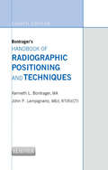
Bontrager's Handbook of Radiographic Positioning and Techniques - E-BOOK
by Kenneth L. Bontrager John Lampignano"The various components contained in this handbook are presented in seamless combination and with a clarity becoming of a much larger work. The book is worthy of recommendation for all those interested in the strenghtening and honing of their core radiographic skills." Reviewed by: RAD Magazine, Barry K Denton, acting radiology services manager, Hywel Dda University Health Board, Wales Date: July 2014
Bontrager's Handbook of Radiographic Positioning and Techniques - E-BOOK
by Kenneth L. Bontrager John LampignanoThis pocket-sized Handbook for Lampignano and Kendrick’s text has it all: new radiographic images, revised critiques, and more. Bontrager’s Handbook of Radiographic Positioning and Techniques, 9th Edition provides bulleted instructions, along with photos of properly positioned patients, to help you safely and confidently position for the most-commonly requested radiographic studies. Suggested techniques and critique points offer a quick reference for evaluating your own radiographs, making it an invaluable tool for learning radiographic positioning in clinical settings.Positioning chapters organized with one projection per page to present a snapshot of information in an easily accessible and portable format. Unique page layout — positioning photos and radiographic images are presented on the same page with the text explanation of each procedure — to show you how the patient should be positioned and what the image should look like. Page number references for the text are included at the bottom of each positioning page so you can easily refer to the text for greater detail and explanation concerning a particular position. 217 projections/positions and 4 conversion charts provide the essential information needed for quick reference. Positioning presentations include positioning instructions, as well as: Collimation guidelines for each projection. Suggested starting exposure factors, including kVp, mAs, SID (source-image receptor distance), type and speed of film and screens, use of grids, and large or small focal spot. Suggested AEC (automatic exposure control) pick-up cell location when photo-timed equipment can be used. Space for writing in exposure factors (techniques) for specific equipment being used. This quick review of information before beginning a procedure helps assure you that the exam is being correctly performed with the least possible patient dose. Appendices offer additional quick-reference information on patient dose, abbreviations and acronyms, and various conversion charts, enabling you to locate important information quickly. NEW! Technique chart updates reflect the latest recommendations for computed and digital radiography.UPDATED! New positioning photos reflect the latest equipment and demonstrate proper positioning. UPDATED! New radiographic images and revised critiques provide examples using the latest technology, and ensure that you are ready to evaluate your own images.EXPANDED! New position added on Apical AP axial give you information and photographs on this position.

Bontrager's Handbook of Radiographic Positioning and Techniques - E-Book: Bontrager's Handbook of Radiographic Positioning and Techniques - E-Book
by John Lampignano Leslie E. KendrickUse this pocket reference in the clinical setting to ensure proper positioning and correct exposure! Handbook of Radiographic Positioning and Techniques, 11th Edition guides you through each step in performing hundreds of imaging procedures. Each projection includes patient positioning photos and concise descriptions to help you produce clear images safely and confidently. Written by the same authors as Textbook of Radiographic Positioning and Related Anatomy, this handbook is a convenient clinical companion to the textbook. A quick review ensures that you perform radiographic procedures right every time! - 297 projections provide a snapshot of essential information in an easily accessible and portable format. - Standard, well-positioned radiograph of each projection demonstrates the critical anatomy that must be visualized, along with a list of evaluation criteria. - Recommendations for radiation protection are provided for each projection, including instructions for shielding. - Positioning pages include a patient position description, recommended automatic exposure control chamber, collimation field size, IR size, CR location and CR angle, suggested kVp ranges, space to fill in exposure factors, and more. - Page number references for the textbook are included at the bottom of each positioning page to easily refer to the text for greater detail and explanation concerning a particular position. - Useful appendices make it easy to find information on patient dose safety, abbreviations and acronyms, and various conversion charts. - NEW! Updated photographs visually demonstrate the latest digital technology used in radiography with new radiographs as well as images of positioning and new equipment. - NEW! The latest ARRT® content specifications and ASRT curriculum guidelines prepare you for certification exams and for clinical practice. - NEW! Additional new projections include a false and modified false profile of the hip, PA clenching wrist for the scapulolunate region, and weight-bearing calcaneus which are performed frequently in orthopedic and sports medicine clinics

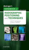
Bontrager's Handbook of Radiographic Positioning and Techniques - E-BOOK: Bontrager's Handbook of Radiographic Positioning and Techniques - E-BOOK
by John Lampignano Leslie E. KendrickGet on-the-spot guidance for all the types of positioning you'll need to perform during clinicals with Bontrager's Handbook of Radiographic Positioning and Techniques, 10th Edition. With bulleted instructions and photos of properly-positioned patients, this portable and pocket-sized reference can help you safely, quickly, and confidently position for the most-commonly requested radiographic studies. Plus, this must-have radiographic positioning and anatomy handbook also provides suggested techniques and critique points to help you quickly and easily evaluate your own radiographs as you produce them in clinicals. - 217 projections provide a snapshot of essential information in an easily accessible and portable format. - Standard radiographic image and evaluation criteria are presented on each positioning page, demonstrating critical anatomy and a list for critique. - Page number references for the text are included at the bottom of each positioning page to help you easily move back and forth between the text for greater detail and explanation concerning a particular position. - Positioning presentations include positioning instructions, collimation field size, CR location and CR angle, suggested kVp ranges, space for writing in exposure factors, and more. - Appendices offer additional quick-reference information on patient dose, abbreviations and acronyms, and various conversion charts, enabling you to locate important information quickly. - NEW! Updated photographs visually demonstrate the latest digital technology used in radiography with new radiographs, positioning, and equipment images. - NEW! Updated content reflecting the latest ARRT competencies prepares you for boards and clinical practice. - NEW! Additional Bernageau and Zanca projections offer guidance on these important projections performed for shoulder pathology and trauma.

Bontrager's Textbook of Radiographic Positioning and Related Anatomy - E-Book
by John Lampignano Leslie E. KendrickMaster radiographic positioning with this comprehensive, user-friendly text. Focusing on one projection per page, Bontrager’s Textbook of Radiographic Positioning and Related Anatomy, 9th Edition includes all of the positioning and projection information you need to know in a clear, bulleted format. Positioning photos, radiographic images, and radiographic overlays, presented side-by-side with the explanation of each procedure, show you how to visualize anatomy and produce the most accurate images. Updated to reflect the latest ARRT competencies and ASRT curriculum guidelines, it features more than 200 of the most commonly requested projections to prepare you for clinical practice.Labeled radiographs (radiographic overlays) identify key radiographic anatomy and landmarks to help you recognize anatomy and determine if you have captured the correct diagnostic information on your images.Positioning chapters, organized with one projection per page, present a manageable amount of information in an easily accessible format. Unique page layout with positioning photos, radiographic images, and radiographic overlays presented side-by-side with the text explanation of each procedure to facilitate comprehension and retention. Pathologic Indications list and define the pathologies most likely to be encountered during procedures covered in each chapter to help you understand the whole patient and improve your ability to produce radiographs that make diagnosis easy for the physician. Pathology Demonstrated sections explain why a particular projection is needed, or what pathology might be demonstrated, to give you a larger frame of reference and a better understanding of the reasoning behind each projection. Radiographic Criteria on positioning pages provide standards for evaluating the quality of each radiograph, helping you develop a routine for evaluating radiographic quality.Pediatric Applications prepare students for clinical success — and prepare technologists to deal competently with the special needs of their pediatric patients.Geriatric Applications include general information on positioning techniques and patient handling for geriatric patients, fostering an understanding of the challenges these patients present to the technologist.Critique Radiographs demonstrate positioning errors and help you avoid similar errors in clinicals. Instructor resources include an accompanying Evolve website with PowerPoint slides, an image collection, and a test bank to help instructors prepare for class.Student resources include a workbook and handbook to help you better understand and retain complicated material.

Bontrager's Textbook of Radiographic Positioning and Related Anatomy - E-Book: Bontrager's Textbook of Radiographic Positioning and Related Anatomy - E-Book
by John Lampignano Leslie E. KendrickGet the information and guidance you need to become proficient in positioning with Bontrager's Textbook of Radiographic Positioning and Related Anatomy, 10th Edition. With a very easy-to-follow organization, this comprehensive text focuses on nearly 200 of the most commonly requested projections to ensure you master what's expected of an entry-level practitioner. And with Bontrager's user-friendly format featuring one projection per page — with bulleted information on the left side of the page and positioning photos, radiographic images, and anatomical drawings aligned on the right — you'll be able to quickly and easily visualize anatomy and master positioning. - Labeled radiographs (radiographic overlays) identify key radiographic anatomy and landmarks to help students recognize anatomy and determine if they have captured the correct diagnostic information on images. - Positioning chapters organized with one projection per page present a manageable amount of information in an easily accessible format. - Unique page layout with positioning photos, radiographic images, and radiographic overlays is presented side-by-side with the text explanation of each procedure to facilitate comprehension and retention. - Clinical Indications features list and define pathologies most likely to be encountered during procedures to help students understand the whole patient and improve their ability to produce radiographs that make diagnosis easy for the physician. - Evaluation Criteria content on positioning pages describes the evaluation/critique process that should be completed for each radiographic image. - Pediatric, Geriatric, and Bariatric Patient Considerations are provided to prepare technologists to accommodate unique patient needs. - Emphasis on radiation safety practices provides recommendations important for clinical practice. - NEW! Updated photographs visually demonstrate the latest digital technology used in radiography with new radiographs, positioning, and equipment images. - UPDATED! The latest ARRT competencies and ASRT curriculum guidelines are incorporated to prepare students for boards and clinical practice. - NEW! Erect positions have been added throughout the text to reflect current practice. - NEW! New Bernageau and Zanca projections have been included to keep students on top of these projections performed for shoulder pathology and trauma. - UPDATED! Critique section at the end of chapters tests students' understanding of common positioning and technical errors found in radiographs. Answer keys are provided for instructors on the Evolve website. - UPDATED! Expanded content on fluoroscopy has been included to keep students up to date on the latest information.
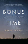
Bonus Time: A true story of surviving the worst and discovering the magic of every day
by Brian PennieHow would you live differently if life gave you a second chance?Brian Pennie shouldn’t be alive today. His drug addiction was so bad that he was deemed too much of a risk for detox. Determined to confront his demons, he went cold turkey at home. Discovered in a pool of blood, it didn’t exactly go to plan, but that’s where his life truly began.On 8 October 2013, he was finally clean after fifteen years of chronic heroin addiction, and something extraordinary happened: the world suddenly became beautiful.Free of the anxiety and fear that had always plagued him, Brian was given a second chance at life, and he devoured every minute of it. Bit by bit he rebuilt his world and began to share what he had learned with others.In this incredibly honest and inspirational book, Brian tells the story of how he turned a seemingly hopeless existence into a rich and rewarding life, showing that change is always possible, no matter how stuck we feel.