- Table View
- List View
Atlas of Esophageal Surgery
by P. Marco Fisichella Marco G. PattiThis Atlas focuses on the description of approaches and surgical techniques used to treat the entire spectrum of esophageal diseases. Surgical “pearls” and tips on how to select and perform the correct operation are included and based both on evidence-based data and the experience of the Editors. Step-by-step descriptions of 14 operative procedures in esophageal surgery are provided. Each chapter describes the current indications, perioperative management strategies, and a detailed operative approach with relevant technical considerations. The description of approaches and surgical techniques used in esophageal surgery are outlined in an easily understandable manner for the specific target audience.
Atlas of Esophageal Surgery
by Fernando A. M. Herbella Marco G. PattiThis Atlas focuses on surgical techniques used to treat the entire spectrum of esophageal diseases. Surgical “pearls” and tips on how to select and perform the correct operation are included and based both on evidence-based data and the experience of the Editors.Step-by-step descriptions of operative and endoscopic procedures in esophageal surgery are provided. Each chapter describes the current indications, perioperative management strategies, and a detailed operative approach with relevant technical considerations.The description of approaches and surgical techniques used in esophageal surgery are outlined in an easily understandable manner for the specific target audience. This book is written by world-class internationally renowned esophageal surgeons and gastroenterologists
Atlas of Esophagus and Stomach Pathology (Atlas of Anatomic Pathology)
by Scott R. Owens Henry D. AppelmanAtlas of Esophagus and Stomach Pathology provides an image-based resource for those studying normal histology of the upper gastrointestinal tract, as well as the microscopic manifestations of developmental abnormalities, toxic insults, infectious diseases, inflammatory and autoimmune conditions, and neoplasia in the esophagus and stomach. Because modern gastrointestinal pathology practice centers on specimens obtained during endoscopic examination, the atlas focuses on biopsy pathology, providing “real-world” microscopic images and ancillary diagnostic studies for most commonly-encountered abnormalities and diseases affecting these two organs. The book is supplemented with endoscopic and special study images. Authored by nationally and internationally recognized pathologists, Atlas of Esophagus and Stomach Pathology is a valuable tool for both pathologists-in-training seeking to make “new acquaintances”, and practicing surgical pathologists in need of a quick visual reference in recalling “old friends” in the world of diagnostic gastrointestinal pathology.
Atlas of Essential Dermatopathology
by Kasia S. Masterpol Andrea Primiani Lyn M. DuncanThe book is not intended to be an all-encompassing atlas or textbook but rather a foundation of principles in dermatopathology, highlighting key elements in the field for trainees and will also serve as a basic resource for the pathologist in general practice. In addition to the sketches and minimal text, we envision accompanying high resolution histopathologic micrographs for ultimate correlation as well.
Atlas of Essential Procedures E-Book: Expert Consult - Online and Print
by Michael Tuggy Jorge GarciaAtlas of Essential Procedures, by Michael Tuggy, MD and Jorge Garcia, MD, makes it easy to perfect the 52 procedures most commonly performed in the primary care setting. Videos and step-by-step illustrations show you how to avoid complications and obtain the best results, and full-text online access makes it easy to reference this title from any computer.Master 52 essential techniques spanning all areas of your practice with separate sections on Dermatology, Obstetrics, Women’s Health, Ultrasound, Urgent/Hospital Care, Gastroenterology, and Musculoskeletal. See what to do step by step with the aid of plentiful full-color illustrations accompanied by clear, practical captions. Observe real-world applications by watching online video demonstrations of 50 procedures including cryosurgery, electrosurgery, catheter placement, circumcision, and shoulder dislocation reduction as well as several basic ob/gyn procedures. Rapidly and conveniently reference the complete contents online at www.expertconsult.com.
Atlas of Exocrine Pancreatic Tumors: Morphology, Biology, and Diagnosis with an International Guide for Tumor Classification
by Daniel S. Longnecker GünterKlöppel YoichiKonishi Parviz M. PourThe classification of tumors is important for understanding tumor histogenesis, for predicting prognosis, for differential diagnosis, and for recommending appropriate therapy. Since 1836, when pancreatic cancer was first described, progress has been made in pancreatic cancer morphology, and a number of classifications have been proposed. All of these classifications are mainly based on morphological characteristics. Some are too detailed to be of practical use while others are more pragmatic. Some of the inherent problems in the previous classifications included difficulties in obtaining an adequate number of pan creatic tumors for examination and insufficient clinical data and follow-up. With the increasing incidence of pancreatic cancer in many parts of the world during the past six decades, and with the availability of more tumors to patho logists, advances have been made in pancreatic tumor studies. Classifications by Cubilla and Fitzgerald and by Kloppel, which are generally similar, mostly considered prominent morphological features and their histogenesis. These pathology-oriented classifications, although complete, were not practical from the standpoint of clinicians concerned with the prognosis of individual tumors.
Atlas of Experimental Toxicological Pathology (Current Histopathology #13)
by C. Gopinath D. Prentice D.J. LewisOur aim in producing a colour atlas of toxicological guidelines itemize the investigations to be carried out pathology was to present a catalogue of histopathologi during the course of the study and they normally include: cal lesions which we had encountered over the years in clinical observations and behaviour; food intake and body various laboratory animal species exposed to a vast weight measurements; serum biochemistry; haema range of pharmaceuticals, agrochemicals and industrial tology; ECG and ophthalmology. At the end of a study, chemicals. While we believe a colour atlas is the ideal full macroscopic and microscopic examinations of the way to share our experiences with others, it quickly organ weight analyses together with tissues are essen became clear to us that for the atlas to be meaningful tial. By far the greater part of the material used in this the associated text must be comprehensive and contain book is from toxicity studies conducted in recent years ample literature references. and performed in compliance with the Good Laboratory The atlas is intended for both the trainee and the Practice standards of governmental regulatory bodies in experienced toxicological pathologist working with lab Europe, Japan and North America. oratory animals in the pharmaceutical, agrochemical or Toxicity studies are commonly carried out in rats, chemical environment.
Atlas of Extreme Facial Cancer: Challenges and Solutions
by Ian Burton Michael F. KlaassenThis book shares an expert experience in managing difficult facial skin cancer and presents rare cases from the authors's own practice. The content follows an anatomical approach based on the complex and often staged reconstruction of extreme facial skin cancer. Written in a clear and logical format, it is intended as a guideline atlas and surgical handbook, defining the principles of CLEAR (Complete Local Excision of the cancer + Aesthetic Reconstruction) and DRAPE (Delayed Reconstruction After Pathological Examination) for reconstructing the face. The book incorporates a wealth of multi-disciplinary knowledge from surgeons, anaesthetists, research scientists, speech / swallowing therapists, pathologists, radiologists and prosthetic rehabilitation specialists. The surgical techniques presented here require a high degree of expertise. Accordingly, the book will be of interest to professionals in the fields of Plastic and Oral and Maxillofacial Surgery, as well as Dermatology.
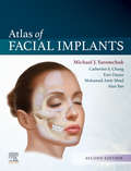
Atlas of Facial Implants E-Book
by Michael J. YaremchukOver the past decade, tremendous innovations in technology, clinical applications, and implant design have transformed the field of facial implant surgery, leading to improved outcomes and greater patient satisfaction. The highly anticipated 2nd Edition of Atlas of Facial Implants, led by renowned plastic surgeon Dr. Michael J. Yaremchuk, brings you fully up to date with these changes, offering authoritative coverage of both aesthetic and reconstructive applications of alloplastic implants for recontouring the craniofacial skeleton.Provides step-by-step descriptions of each procedure enhanced by hundreds of color illustrations and color photographs depicting preoperative, intraoperative, and postoperative views. Reviews indications for implant use, patient evaluation, and surgical planning, as well as pearls and pitfalls throughout. Discusses Computer-Aided Design (CAD)/Computed-Aided Manufacture (CAM) for both cranial reconstruction (cranioplasty) as well as aesthetic applications. Features new coverage of facial implants as an important adjunct in rejuvenative aesthetic surgery, as well as their role in refining orthognathic surgical procedures, surgical treatment of Graves’ disease, and facial skeletal augmentation. Provides access to procedural videos depicting post orthognathic irregularities and imbalances, functional cranioplasty, and more.
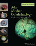
Atlas of Feline Ophthalmology
by Kerry L. Ketring Mary Belle GlazeSuccessful management of eye disease relies on the veterinarian’s ability to identify ocular features and distinguish pathologic changes. Atlas of Feline Ophthalmology, Second Edition is an invaluable diagnostic reference, providing high-quality color photographs for comparison with a presenting complaint. Presenting 394 photographs illustrating both normal and pathologic ocular conditions, this Second Edition offers a current, complete reference on ocular diseases, adding conditions recognized since publication of the first edition, a broader geographic scope, and many new images with improved quality. Carefully designed for easy reference, the contents are divided into sections corresponding to specific anatomical structures of the eye. A useful appendix new to this edition groups figures by etiology, making it easy to find every image associated with a specific agent or disease. Atlas of Feline Ophthalmology, Second Edition is a useful tool aiding general practitioners in diagnosing eye disease in cats.
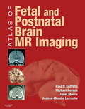
Atlas of Fetal and Postnatal Brain MR
by Paul D. Griffiths Janet Morris Jeanne-Claudie Larroche Michael ReevesThe Atlas of Fetal and Neonatal Brain MR is an excellent atlas that fills the gap in coverage on normal brain development. Dr. Paul Griffiths and his team present a highly visual approach to the neonatal and fetal periods of growth. With over 800 images, you’ll have multiple views of normal presentation in utero, post-mortem, and more. Whether you’re a new resident or a seasoned practitioner, this is an invaluable guide to the new and increased use of MRI in evaluating normal and abnormal fetal and neonatal brain development.Covers both fetal and neonatal periods to serve as the most comprehensive atlas on the topic. Features over 800 images for a focused visual approach to applying the latest imaging techniques in evaluating normal brain development. Presents multiple image views of normal presentation to include in utero and post-mortem images (from coronal, axial, and sagittal planes), gross pathology, and line drawings for each gestation.
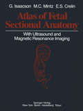
Atlas of Fetal Sectional Anatomy: With Ultrasound and Magnetic Resonance Imaging
by Glenn Isaacson Marshall C. Mintz Edmund S. CrelinThe fetal period of human growth and development has become an area of intense study in recent years, due in large part to the development of diagnostic ultrasound. More than 2,000 articles have been published in the last five years describing anatomy and pathology in utero, as reflected in sonographic images. Yet, no stan dard reference exists to correlate these images with fetal gross anatomy and at tempts to draw parallels from adult structure have often led to false assumptions. The dictum "the newborn is not a miniature adult" is all the more valid for the fetus. This text aims to provide a comprehensive reference for normal sectional anat omy correlated with in utero ultrasound images. In addition, magnetic resonance images of therapeutically aborted or stillborn fetuses are paired with similar gross sections to serve as a foundation upon which current in vivo studies may build. Lastly, a miscellaneous section illustrates several anatomic points useful in the understanding of fetal anatomy. These points include the changing anatomy of the fetal brain during gestation and the anatomy of the meninges, the fetal heart, and ductus venosus. It is our hope that this atlas will provide a clear picture of fetal anatomy, rectify some of the confusion which exists in antenatal diagnosis, and stimulate further interest in fetal development.
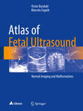
Atlas of Fetal Ultrasound: Normal Imaging and Malformations
by Victor Bunduki Marcelo ZugaibThe present book is an illustrated guide on fetal medicine, including a wealth of normal and pathological/malformations ultrasound images, throughout the whole pregnancy. Thus, the book intends to fill two gaps in once: The lack of a material discussing the basic principles of fetal ultrasound, which are the basement for a more efficient learning in fetal medicine.. The need for a thorough approach in fetal medicine, presenting both normal and pathological imaging, allowing a detailed evaluation of clinical conditions of importance in prenatal care and follow up. The Atlas of Ultrasound Imaging is an up to date guide to all obstetricians, gynecologists and pediatricians who intend to upgrade their knowledge in fetal medicine, as well as to any other professional, professor, student or research interested in fetal ultrasound.
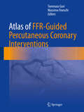
Atlas of FFR-Guided Percutaneous Coronary Interventions
by Tommaso Gori Massimo FineschiThis book reviews the theory and practice of fractional flow reserve (FFR)-guided coronary intervention, a technique that gives sense and a rationale to daily decisions in the interventional suite. FFR guidance provides detailed information on coronary hemodynamics for the interventional cardiologist. This technique has profound practical implications for therapeutic decisions and for the prognosis of patients. The Atlas of FFR-Guided Percutaneous Coronary Interventions provides practicing physicians clear information to understand both the complexity of the technique and the correct way to apply it. It is designed both to assist younger faculty and those in training, and to act as a clinical resource for more experienced practitioners. Using the clinical cases outlined, the reader can learn to appreciate the pitfalls, tips and tricks that simplify the performance and interpretation of FFR and iFR.
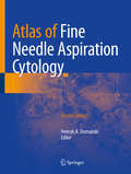
Atlas of Fine Needle Aspiration Cytology
by Henryk A. DomanskiThis updated and expanded second edition covers all of the diagnostic areas where FNAC is used today. Each chapter follows a similar, practical format: diagnostic criteria with an emphasis of differential diagnoses; diagnostic problems and pitfalls; and relevant findings of ancillary methods. Authoritative discussions will reflect accepted international viewpoints. The interaction of the cytologist or cytopathologist with other specialists (radiologists, oncologists and surgeons) is emphasized and illustrated throughout. With contributions from experts in the field internationally and many new colour images Atlas of Fine Needle Aspiration Cytology, Second Edition provides a comprehensive and up-to-date guide to FNAC for pathologists, cytopathologists, radiologists, oncologists, surgeons and others involved in the diagnosis and treatment of patients with suspicious mass lesions.
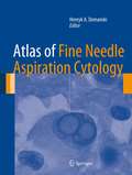
Atlas of Fine Needle Aspiration Cytology
by Henryk A. DomanskiThis book covers all of the diagnostic areas where FNAC is used today. This includes palpable lesions and lesions sampled using various radiological methods, and correlations with ancillary examinations detailed on an entity-by-entity basis. As well as being a complete atlas of the facts and findings important to FNAC, this atlas is a guide to diagnostic methods that optimize health care. The interaction of the cytologist or cytopathologist with other specialists (radiologists, oncologists and surgeons) involved in the diagnosis and treatment of patients with suspicious mass lesions is emphasized and illustrated throughout. With contributions from experts in the field internationally and abundant colour images Atlas of Fine Needle Aspiration Cytology provides a comprehensive and up-to-date guide to FNAC for pathologists, cytopathologists, radiologists, oncologists, surgeons and others involved in the diagnosis and treatment of patients with suspicious mass lesions.
Atlas of Finger Reconstruction: Techniques and Cases
by Jian Lin Jianli Wang Deqing Hu Yongqing Xu Tianhao ZhangThis book covers latest advancement of finger reconstruction caused by severe injury leading to finger defects, presenting amount of valuable clinical experience and research achievement. This book provides practical guidance for hand and foot surgeons, micro- and reconstructive surgeons, trauma surgeons, orthopedic surgeons, general practitioners as well as trainees.The book mainly contains two parts: Part 1 focuses on development of the history of finger reconstruction, applied anatomy of the extremities, commonly used equipment and materials, frequently used medicines, preoperative treatment, selection of anesthesia, fundamental skill for finger reconstruction, postoperative treatment and management and methods of functional recovery after finger reconstruction. It is not uncommon in clinical scenarios that severe traumas caused by high energy damage in hands leading to life-long disability. The initial injury management is crucial. Surgeons face the arduous task of attempting to repair and reconstruct the injuries, reduce the patient’s disability and improve their quality of life. Part 2 demonstrates the concepts of different types of finger reconstruction with key points described by case presentations. Depending on the types and the severity of the lesions, accuracy of doctor’s judgment and proficiency of surgery skill have vital significance during the treatment of hand trauma together with patients’ compliance on functional rehabilitation.
Atlas of Forensic and Criminal Psychology
by Bernat-N. TiffonOriginally published in Spanish in 2017 by Libreria Bosch, Barcelona, the Atlas of Forensic and Criminal Psychology is a one-of-kind book made available in English for the first time. This unique work is highly illustrated with full-color images, providing a medico-legal examination of forensic pathology as it relates to cases of forensic psychological interest. The book begins with a historical perspective and includes images of patients to familiarize the reader with symptoms, the hazard-risk criteria, lethality, and suicidal rescue—research that Dr. Tiffon has addressed in his previous publications. Chapters present photographic records of cases to deepen forensic, psychologist, and medico-legal professionals’ insight into thoughts, behaviors, and mechanisms of self- and hetero-aggressiveness. Such cases illustrate the outcomes of various disorders manifested in individuals and victims; as such, they provide an understanding of the psychological-legal conclusions reached in such cases in order to adapt the legal and preventative measures for specific situations. Coverage includes affective, schizophrenic, and personality disorders as contributing elements in diagnostic judgments, noting the great difficulty such examples present to experts performing psychopathological evaluations after criminal, and often violent, events have occurred. Various psychopathological disorders are addressed as well as the technical treatment that should occur in each case from a psychological-forensic perspective. Features: • Presents a provocative look at various syndromes familiar to forensic psychologists, as applied to criminal cases and the pathology of suicide victims and homicide perpetrators • Combines the work of world-renowned expert contributors to examine the criminal, legal, and psychological facets of various diagnoses and case examples • Offers insight into the psychological state of suicide victims, considering their state of mind as a "psychological autopsy" In his previous books published in Spanish, Manual of Consulting in Psychology and Clinical, Legal, Legal, Criminal, and Forensic Psychopathology (2008), Manual of Professional Performance in Clinical, Criminal, and Forensic Psychopathology (2009), and the 4-volume Practical Criminological Atlas of Forensic Psychometry (2019-2020), Tiffon approached forensic psychology and psychopathology from a theoretical perspective. In the Atlas of Forensic and Criminal Psychology, his first book translated into English, Tiffon expands on these prior works, serving to provide a visual reference and guide to medical pathologists and consulting psychologists in cases of disorders in which psychopathological mutilation, injury, and self-injury occur.
Atlas of Forensic Pathology: For Police, Forensic Scientists, Attorneys, And Death Investigators (pdf)
by Joseph A. Prahlow and Roger W. Byard
Atlas of Frontal Sinus Surgery: A Comprehensive Surgical Guide
by David R. Lobo Jaime Viera Artiles Javier A. OspinaThe atlas offers a comprehensive and up-to-date overview of frontal sinus surgery. In recent years there have been great advances in endoscopic nasosinusal surgery but they have been particularly prominent in frontal sinus surgery. The book provides complete instructions for a gradual learning of the different surgical techniques and includes surgical pearls. It is enriched with videos presenting real-time guidance for frontal sinus endoscopic procedures. The book will meet the needs of both trainees and more experienced practitioners, and will enable them to make steady progress in endoscopic surgery and to adopt a more complete and safe approach to the frontal sinus. It will be of interest also for ophthalmologists, maxillofacial surgeons and neurosurgeons.
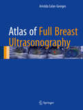
Atlas of Full Breast Ultrasonography
by Aristida Colan-GeorgesThis atlas describes and illustrates a novel approach, referred to as full breast ultrasonography (FBU), that represents a challenge to conventional breast imaging diagnosis. The coverage encompasses examination technique, diagnostic criteria, the imaging features of a wide variety of lesions, and role in follow-up. FBU involves anatomic ultrasound scanning based on the ductal echography technique proposed by Michel Teboul, supplemented by Doppler and real-time sonoelastography. The approach offers a variety of advantages. Compared with MRI it has a lower cost, wider availability, better resolution, and improved correlation with anatomy. Compared with mammography it has the benefits of absence of irradiation and pain, applicability in all cases, and better overall accuracy. Furthermore, the standardized technique of acquisition and interpretation means that it is suitable as a screening test, unlike classic ultrasonography. FBU is applicable in ultrasound BI-RADS assessment and is of value in depicting both benign and malignant conditions. It can be recommended as a first-line method of diagnosis and for the follow-up of treated breasts, regardless of the patient’s age, sex, or physical condition.
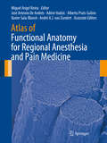
Atlas of Functional Anatomy for Regional Anesthesia and Pain Medicine: Human Structure, Ultrastructure and 3D Reconstruction Images
by Miguel Angel Reina José Antonio De Andrés Admir Hadzic Alberto Prats-Galino Xavier Sala-Blanch André A.J. van ZundertThis is the first atlas to depict in high-resolution images the fine structure of the spinal canal, the nervous plexuses, and the peripheral nerves in relation to clinical practice. The Atlas of Functional Anatomy for Regional Anesthesia and Pain Medicine contains more than 1500 images of unsurpassed quality, most of which have never been published, including scanning electron microscopy images of neuronal ultrastructures, macroscopic sectional anatomy, and three-dimensional images reconstructed from patient imaging studies. Each chapter begins with a short introduction on the covered subject but then allows the images to embody the rest of the work; detailed text accompanies figures to guide readers through anatomy, providing evidence-based, clinically relevant information. Beyond clinically relevant anatomy, the book features regional anesthesia equipment (needles, catheters, surgical gloves) and overview of some cutting edge research instruments (e.g. scanning electron microscopy and transmission electron microscopy). Of interest to regional anesthesiologists, interventional pain physicians, and surgeons, this compendium is meant to complement texts that do not have this type of graphic material in the subjects of regional anesthesia, interventional pain management, and surgical techniques of the spine or peripheral nerves.
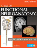
Atlas of Functional Neuroanatomy
by Walter J. HendelmanUnderstanding how the brain is organized and visualizing its pathways and connections can be conceptually challenging. The Atlas of Functional Neuroanatomy, Third Edition addresses this challenge by presenting a clear visual guide to the human central nervous system (CNS). This edition has been completely reorganized to facilitate learning the stru