- Table View
- List View

Atlas of Endovascular Venous Surgery E-Book
by Jose AlmeidaHighly visual and packed with useful, practical information, Atlas of Endovascular Venous Surgery, 2nd Edition, provides real-world instruction on the evaluation, diagnostic imaging, and medical and endovascular surgical management of acute and chronic venous diseases. Dr. Jose Almeida, pioneering expert in the field and host of the annual International Vein Congress, along with other highly regarded practitioners, offers an authoritative understanding of what causes increased venous pressure and solutions for reducing venous hypertension. Detailed, full-color intraoperative illustrations capture key teaching moments, helping you better understand the nuances of surgery and improve your ability to perform cutting-edge procedures.
Atlas of Enteroscopy: Endoscopy of the Small and Large Bowel; Retrograde Cholangio-Pancreatography
by L. Demling M. Classen P. FruehmorgenIt is my pleasure to introduce you to this new Atlas by Professor DEMLING and his colleagues on the very timely subjects colonoscopy, duodenoscopy and endoscopic cannu lation of the bile and pancreatic ducts. Professor DEMLING, unlike many others who carry his teaching and administra tive burdens, continues to be very personally involved in performing endoscopies. His clinic is one of the best organized and best equipped in the world. Professor DEM LING and his colleagues have been instrumental in introducing to the European continent these new techniques of duodeno scopy, colonoscopy and bile duct cannulation which were originally developed in Japan. They have added significant contributions of their own and now present to the reader a clear, concise, very well illustrated description of these methods. Dr. CLASSEN is one of the pioneers in endoscopic cannula tion. He has been kind enough to come to the United States and share his expertise with us at several Post Graduate Education programs. The students in these courses have been most enthusiastic about his presentations. Through this text he makes his extensive experience available to all endo scopists. The beginner at cannulation will find the illustrations of the various shapes the papilla may assume most helpful. I had the pleasure this summer of teaching a course in Brazil with Dr. FRUHMORGEN. Very few physicians have con centrated on colonoscopy to the extent that he has.
Atlas of Enteroscopy
by Gerard J. Gay, Francesco P. RossiniIn recent years, important technological innovations have made it possible to evaluate the small bowel endoscopically. Thus, traditional diagnostic methods such as imaging techniques are flanked by enteroscopy. The aim of this book is to present the results of enteroscopy as a method to diagnose small bowel disease, correlating it to the other investigative methods and above all to clinical findings. The book is a unique selected collection of images deriving from the results of documented experience and also including a brief clinical presentation, tables and diagrams of diagnostic algorithms. Renowned and accredited experts in gastroenterology, enteroscopy and researchers in the sector of small bowel disease have contributed to this volume.
Atlas Of Epilepsies
by C. P. Panayiotopoulos S. R. Benbadis R. G. Beran A. T. Berg J. Engel A. S. Galanopoulou P. W. Kaplan M. Koutroumanidis S. L. Moshe D. R. Nordli J. M. Serratosa S. M. Sisodiya W. O. Tatum T. Valeta A. Wilner
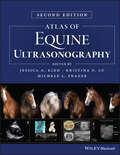
Atlas of Equine Ultrasonography
by Jessica A. Kidd Kristina G. Lu Michele L. FrazerThe only visual guide to equine ultrasonography based on digital ultrasound technology. Atlas of Equine Ultrasonography provides comprehensive coverage of both musculoskeletal and non-musculoskeletal areas of the horse. Ideal for practitioners in first opinion or referral practices, each chapter features normal images for anatomical reference followed by abnormal images covering a broad range of recognised pathologies. The book is divided into musculoskeletal, reproductive and internal medicine sections and includes positioning diagrams demonstrating how to capture optimal images. With contributions from experts around the world, this book is the go-to reference for equine clinical ultrasonography. Key features include: Pictorially based with a wealth of digital ultrasound images covering both musculoskeletal and non-musculoskeletal areas and their associated pathologies. Each chapter begins with a discussion of normal anatomy and demonstrates how to obtain and interpret the images presented. A video library of over 50 ultrasound examinations is available for streaming or download and viewing on-the-go. Access details are provided in the book.
Atlas of Equine Ultrasonography
by Jessica A. Kidd Kristina G. Lu Michele L. FrazerATLAS OF EQUINE ULTRASONOGRAPHY A THOROUGH EXPANSION TO THE FIRST ATLAS OF ULTRASONOGRAPHY IN THE HORSE, WITH NEW AND SIGNIFICANTLY IMPROVED IMAGES Ultrasonography is a vital diagnostic tool that can be applied in numerous functions in a veterinary practice. In conjunction with relevant clinical information—patient history and physical examination findings, for example—it can act as an important aid in the veterinarian’s decision-making process. Many vets in equine practice rely upon ultrasonography as a mainstay of equine diagnostic imaging on a wide range of structures and body systems. Ultrasonography is a useful procedure that is non-invasive and acts in complement to radiography to successfully diagnose the animal’s condition. This book’s aim is to encourage the clinician to rely further on the use of ultrasonography in their practice. The second edition of Atlas of Equine Ultrasonography provides an updated and expanded revision of the first atlas of ultrasonography in the horse. The first edition of this important resource was the first pictorially-based book to cover ultrasonography in the horse, and remains the only book currently available on the subject. The current version offers 450 additional images with greater clarity and precision in the images throughout and demonstrates how to obtain images in each body region while offering clinical ultrasonograms that show pathology. Atlas of Equine Ultrasonography readers will also find: High-quality clinical ultrasonograms for important musculoskeletal, reproductive, and medical conditions in the horse More than 1,500 images, with accompanying concise text describing the images A companion website that provides video clips showing dynamic ultrasound exams Atlas of Equine Ultrasonography is an invaluable reference to any veterinarian evaluating ultrasonograms in equine patients. As a result, this book will be of particular interest to equine specialists, veterinary radiologists, equine practitioners, and veterinary students.
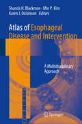
Atlas of Esophageal Disease and Intervention: A Multidisciplinary Approach
by Shanda H. Blackmon Min P. Kim Karen J. DickinsonThis atlas provides a comprehensive, state-of-the-art review of all interventions that pertain to the esophagus. It includes a review of the current staging modalities, ablation technologies, resection and reconstruction techniques, and disease classification. Evidence-based guidelines regarding how each intervention is chosen are also included. With color illustrations and photographs for each surgery, the atlas details specific anatomic topics such as micro-anatomy of Barrett’s and Dysplasia, EMR pathology, endoscopic ultrasound, and conventional surgical anatomy. Each intervention is presented in task format as a task list to be checked-off as each step is completed.Written by experts in the field, Atlas of Esophageal Disease and Intervention: A Multidisciplinary Approach serves as a valuable resource for any practitioner who performs esophageal intervention and will guide new surgeons and gastroenterologists into the hybrid multidisciplinary approach to this disease.
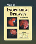
Atlas of Esophageal Diseases
by Roy C. Orlando Mark FeldmanThe updated and expanded Second edition of the Atlas of Esophageal Diseases provides an 11-chapter visual panorama of the esophagus in both health and disease. More than 600 detailed images, including many hand-drawn illustrations, cover all aspects of the esophagus. The Atlas opens with a chapter on esophageal anatomy and physiology, and takes the reader through an exploration of gastroesophageal reflux disease, pH-monitoring, and finally, surgery of the esophagus.
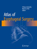
Atlas of Esophageal Surgery
by P. Marco Fisichella Marco G. PattiThis Atlas focuses on the description of approaches and surgical techniques used to treat the entire spectrum of esophageal diseases. Surgical “pearls” and tips on how to select and perform the correct operation are included and based both on evidence-based data and the experience of the Editors. Step-by-step descriptions of 14 operative procedures in esophageal surgery are provided. Each chapter describes the current indications, perioperative management strategies, and a detailed operative approach with relevant technical considerations. The description of approaches and surgical techniques used in esophageal surgery are outlined in an easily understandable manner for the specific target audience.
Atlas of Esophageal Surgery
by Fernando A. M. Herbella Marco G. PattiThis Atlas focuses on surgical techniques used to treat the entire spectrum of esophageal diseases. Surgical “pearls” and tips on how to select and perform the correct operation are included and based both on evidence-based data and the experience of the Editors.Step-by-step descriptions of operative and endoscopic procedures in esophageal surgery are provided. Each chapter describes the current indications, perioperative management strategies, and a detailed operative approach with relevant technical considerations.The description of approaches and surgical techniques used in esophageal surgery are outlined in an easily understandable manner for the specific target audience. This book is written by world-class internationally renowned esophageal surgeons and gastroenterologists
Atlas of Esophagus and Stomach Pathology (Atlas of Anatomic Pathology)
by Scott R. Owens Henry D. AppelmanAtlas of Esophagus and Stomach Pathology provides an image-based resource for those studying normal histology of the upper gastrointestinal tract, as well as the microscopic manifestations of developmental abnormalities, toxic insults, infectious diseases, inflammatory and autoimmune conditions, and neoplasia in the esophagus and stomach. Because modern gastrointestinal pathology practice centers on specimens obtained during endoscopic examination, the atlas focuses on biopsy pathology, providing “real-world” microscopic images and ancillary diagnostic studies for most commonly-encountered abnormalities and diseases affecting these two organs. The book is supplemented with endoscopic and special study images. Authored by nationally and internationally recognized pathologists, Atlas of Esophagus and Stomach Pathology is a valuable tool for both pathologists-in-training seeking to make “new acquaintances”, and practicing surgical pathologists in need of a quick visual reference in recalling “old friends” in the world of diagnostic gastrointestinal pathology.
Atlas of Essential Dermatopathology
by Kasia S. Masterpol Andrea Primiani Lyn M. DuncanThe book is not intended to be an all-encompassing atlas or textbook but rather a foundation of principles in dermatopathology, highlighting key elements in the field for trainees and will also serve as a basic resource for the pathologist in general practice. In addition to the sketches and minimal text, we envision accompanying high resolution histopathologic micrographs for ultimate correlation as well.

Atlas of Essential Procedures E-Book: Expert Consult - Online and Print
by Michael Tuggy Jorge GarciaAtlas of Essential Procedures, by Michael Tuggy, MD and Jorge Garcia, MD, makes it easy to perfect the 52 procedures most commonly performed in the primary care setting. Videos and step-by-step illustrations show you how to avoid complications and obtain the best results, and full-text online access makes it easy to reference this title from any computer. - Master 52 essential techniques spanning all areas of your practice with separate sections on Dermatology, Obstetrics, Women's Health, Ultrasound, Urgent/Hospital Care, Gastroenterology, and Musculoskeletal. - See what to do step by step with the aid of plentiful full-color illustrations accompanied by clear, practical captions. - Observe real-world applications by watching online video demonstrations of 50 procedures including cryosurgery, electrosurgery, catheter placement, circumcision, and shoulder dislocation reduction as well as several basic ob/gyn procedures. - Rapidly and conveniently reference the complete contents online at www.expertconsult.com.
Atlas of Exocrine Pancreatic Tumors: Morphology, Biology, and Diagnosis with an International Guide for Tumor Classification
by Daniel S. Longnecker GünterKlöppel YoichiKonishi Parviz M. PourThe classification of tumors is important for understanding tumor histogenesis, for predicting prognosis, for differential diagnosis, and for recommending appropriate therapy. Since 1836, when pancreatic cancer was first described, progress has been made in pancreatic cancer morphology, and a number of classifications have been proposed. All of these classifications are mainly based on morphological characteristics. Some are too detailed to be of practical use while others are more pragmatic. Some of the inherent problems in the previous classifications included difficulties in obtaining an adequate number of pan creatic tumors for examination and insufficient clinical data and follow-up. With the increasing incidence of pancreatic cancer in many parts of the world during the past six decades, and with the availability of more tumors to patho logists, advances have been made in pancreatic tumor studies. Classifications by Cubilla and Fitzgerald and by Kloppel, which are generally similar, mostly considered prominent morphological features and their histogenesis. These pathology-oriented classifications, although complete, were not practical from the standpoint of clinicians concerned with the prognosis of individual tumors.
Atlas of Experimental Toxicological Pathology (Current Histopathology #13)
by C. Gopinath D. Prentice D.J. LewisOur aim in producing a colour atlas of toxicological guidelines itemize the investigations to be carried out pathology was to present a catalogue of histopathologi during the course of the study and they normally include: cal lesions which we had encountered over the years in clinical observations and behaviour; food intake and body various laboratory animal species exposed to a vast weight measurements; serum biochemistry; haema range of pharmaceuticals, agrochemicals and industrial tology; ECG and ophthalmology. At the end of a study, chemicals. While we believe a colour atlas is the ideal full macroscopic and microscopic examinations of the way to share our experiences with others, it quickly organ weight analyses together with tissues are essen became clear to us that for the atlas to be meaningful tial. By far the greater part of the material used in this the associated text must be comprehensive and contain book is from toxicity studies conducted in recent years ample literature references. and performed in compliance with the Good Laboratory The atlas is intended for both the trainee and the Practice standards of governmental regulatory bodies in experienced toxicological pathologist working with lab Europe, Japan and North America. oratory animals in the pharmaceutical, agrochemical or Toxicity studies are commonly carried out in rats, chemical environment.
Atlas of Extreme Facial Cancer: Challenges and Solutions
by Ian Burton Michael F. KlaassenThis book shares an expert experience in managing difficult facial skin cancer and presents rare cases from the authors's own practice. The content follows an anatomical approach based on the complex and often staged reconstruction of extreme facial skin cancer. Written in a clear and logical format, it is intended as a guideline atlas and surgical handbook, defining the principles of CLEAR (Complete Local Excision of the cancer + Aesthetic Reconstruction) and DRAPE (Delayed Reconstruction After Pathological Examination) for reconstructing the face. The book incorporates a wealth of multi-disciplinary knowledge from surgeons, anaesthetists, research scientists, speech / swallowing therapists, pathologists, radiologists and prosthetic rehabilitation specialists. The surgical techniques presented here require a high degree of expertise. Accordingly, the book will be of interest to professionals in the fields of Plastic and Oral and Maxillofacial Surgery, as well as Dermatology.

Atlas of Facial Implants E-Book
by Michael J. YaremchukOver the past decade, tremendous innovations in technology, clinical applications, and implant design have transformed the field of facial implant surgery, leading to improved outcomes and greater patient satisfaction. The highly anticipated 2nd Edition of Atlas of Facial Implants, led by renowned plastic surgeon Dr. Michael J. Yaremchuk, brings you fully up to date with these changes, offering authoritative coverage of both aesthetic and reconstructive applications of alloplastic implants for recontouring the craniofacial skeleton.Provides step-by-step descriptions of each procedure enhanced by hundreds of color illustrations and color photographs depicting preoperative, intraoperative, and postoperative views. Reviews indications for implant use, patient evaluation, and surgical planning, as well as pearls and pitfalls throughout. Discusses Computer-Aided Design (CAD)/Computed-Aided Manufacture (CAM) for both cranial reconstruction (cranioplasty) as well as aesthetic applications. Features new coverage of facial implants as an important adjunct in rejuvenative aesthetic surgery, as well as their role in refining orthognathic surgical procedures, surgical treatment of Graves’ disease, and facial skeletal augmentation. Provides access to procedural videos depicting post orthognathic irregularities and imbalances, functional cranioplasty, and more.
Atlas of Feline Ophthalmology
by Kerry L. Ketring Mary Belle GlazeSuccessful management of eye disease relies on the veterinarian’s ability to identify ocular features and distinguish pathologic changes. Atlas of Feline Ophthalmology, Second Edition is an invaluable diagnostic reference, providing high-quality color photographs for comparison with a presenting complaint. Presenting 394 photographs illustrating both normal and pathologic ocular conditions, this Second Edition offers a current, complete reference on ocular diseases, adding conditions recognized since publication of the first edition, a broader geographic scope, and many new images with improved quality. Carefully designed for easy reference, the contents are divided into sections corresponding to specific anatomical structures of the eye. A useful appendix new to this edition groups figures by etiology, making it easy to find every image associated with a specific agent or disease. Atlas of Feline Ophthalmology, Second Edition is a useful tool aiding general practitioners in diagnosing eye disease in cats.
Atlas of Fetal and Postnatal Brain MR
by Paul D. Griffiths Janet Morris Jeanne-Claudie Larroche Michael ReevesThe Atlas of Fetal and Neonatal Brain MR is an excellent atlas that fills the gap in coverage on normal brain development. Dr. Paul Griffiths and his team present a highly visual approach to the neonatal and fetal periods of growth. With over 800 images, you’ll have multiple views of normal presentation in utero, post-mortem, and more. Whether you’re a new resident or a seasoned practitioner, this is an invaluable guide to the new and increased use of MRI in evaluating normal and abnormal fetal and neonatal brain development.Covers both fetal and neonatal periods to serve as the most comprehensive atlas on the topic. Features over 800 images for a focused visual approach to applying the latest imaging techniques in evaluating normal brain development. Presents multiple image views of normal presentation to include in utero and post-mortem images (from coronal, axial, and sagittal planes), gross pathology, and line drawings for each gestation.
Atlas of Fetal Sectional Anatomy: With Ultrasound and Magnetic Resonance Imaging
by Glenn Isaacson Marshall C. Mintz Edmund S. CrelinThe fetal period of human growth and development has become an area of intense study in recent years, due in large part to the development of diagnostic ultrasound. More than 2,000 articles have been published in the last five years describing anatomy and pathology in utero, as reflected in sonographic images. Yet, no stan dard reference exists to correlate these images with fetal gross anatomy and at tempts to draw parallels from adult structure have often led to false assumptions. The dictum "the newborn is not a miniature adult" is all the more valid for the fetus. This text aims to provide a comprehensive reference for normal sectional anat omy correlated with in utero ultrasound images. In addition, magnetic resonance images of therapeutically aborted or stillborn fetuses are paired with similar gross sections to serve as a foundation upon which current in vivo studies may build. Lastly, a miscellaneous section illustrates several anatomic points useful in the understanding of fetal anatomy. These points include the changing anatomy of the fetal brain during gestation and the anatomy of the meninges, the fetal heart, and ductus venosus. It is our hope that this atlas will provide a clear picture of fetal anatomy, rectify some of the confusion which exists in antenatal diagnosis, and stimulate further interest in fetal development.
Atlas of Fetal Ultrasound: Normal Imaging and Malformations
by Victor Bunduki Marcelo ZugaibThe present book is an illustrated guide on fetal medicine, including a wealth of normal and pathological/malformations ultrasound images, throughout the whole pregnancy. Thus, the book intends to fill two gaps in once: The lack of a material discussing the basic principles of fetal ultrasound, which are the basement for a more efficient learning in fetal medicine.. The need for a thorough approach in fetal medicine, presenting both normal and pathological imaging, allowing a detailed evaluation of clinical conditions of importance in prenatal care and follow up. The Atlas of Ultrasound Imaging is an up to date guide to all obstetricians, gynecologists and pediatricians who intend to upgrade their knowledge in fetal medicine, as well as to any other professional, professor, student or research interested in fetal ultrasound.
Atlas of FFR-Guided Percutaneous Coronary Interventions
by Tommaso Gori Massimo FineschiThis book reviews the theory and practice of fractional flow reserve (FFR)-guided coronary intervention, a technique that gives sense and a rationale to daily decisions in the interventional suite. FFR guidance provides detailed information on coronary hemodynamics for the interventional cardiologist. This technique has profound practical implications for therapeutic decisions and for the prognosis of patients. The Atlas of FFR-Guided Percutaneous Coronary Interventions provides practicing physicians clear information to understand both the complexity of the technique and the correct way to apply it. It is designed both to assist younger faculty and those in training, and to act as a clinical resource for more experienced practitioners. Using the clinical cases outlined, the reader can learn to appreciate the pitfalls, tips and tricks that simplify the performance and interpretation of FFR and iFR.