- Table View
- List View
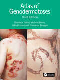
Atlas of Genodermatoses
by Gianluca Tadini Michela Brena Lidia Pezzani Francesca BesagniGenodermatoses are often considered rare diseases seldom seen by practicing clinicians, but as a result, professionals often have little experience or confidence with their diagnosis when they are called upon for a clinical case.This text presents a comprehensive illustrated overview of almost 200 inherited diseases of the skin, hair, and nails. Examples have been expanded, with new images added to provide clear examples, alongside coherent and comprehensive explanations to enable clinicians to easily identify and source relevant information. This resource encompasses a varied range of skin diseases, providing accessible and in-depth information to help familiarise clinicians. The entry for each disease provides background, followed by common characterisations, manifestations, laboratory findings, genetics, cutaneous and extracutaneous findings, differential diagnosis, an overview of complications and recommended follow-ups.Authored by dermatologists and geneticists, this is an atlas of scientific research which updates established information with current studies and references. In its third edition, this text becomes an invaluable resource for dermatologists and pediatricians.
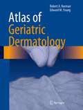
Atlas of Geriatric Dermatology
by Robert A. Norman Edward M. Young, JrThis is a comprehensive, practical, densely illustrated diagnostic and therapeutic guide for all geriatric dermatology providers. The book comprises 50 chapters and over 600 color photographs on topics ranging from common conditions such as basal cell carcinoma, rosacea, and seborrheic dermatitis to unusual conditions such as angiosarcoma, dermatofibrosarcoma protuberans, and porphyria cutanea tarda.Sections include:- Inflammatory conditions (including contact dermatitis, alopecia, erythema multiforme, pemphigus, bullous pemphigoid, porphyria, pruritus, psoriasis, rosacea, seborrhea, urticaria, xerosis, and more)- Infections (fungus, herpes simplex and zoster, scabies, lice, and warts)- Skin signs in systemic disease (skin tags, cutaneous metastases, xanthomas)- Regional dermatoses (intertrigo, leg ulcers, pressure sores)- Benign tumors (chondrodermatitis, cysts, ganglion, fibrous papule, seborrheic keratoses, lentigines, and benign vascular lesions)- Pre-malignant and malignant tumors (actinic keratoses, angiosarcoma, basal cell carcinoma, dermatofibroma and dermatofibrosarcoma protuberans, intraepidermal neoplasia, Kaposi's sarcoma, keratoacanthoma, lentigo maligna, cutaneous lymphoma, Mycosis fiungoides, melanoma, nevi and moles, and squamous cell carcinoma)
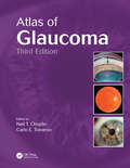
Atlas of Glaucoma
by Neil T. Choplin Carlo E. TraversoGlaucoma affects all age groups and is a leading cause of blindness worldwide. It is imperative that practicing clinicians and surgeons recognize both primary and secondary glaucoma as well as cases of glaucoma associated with other disorders. Atlas of Glaucoma, Third Edition provides an in-depth review and analysis of the management of glaucoma an
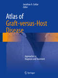
Atlas of Graft-versus-Host Disease: Approaches to Diagnosis and Treatment
by Jonathan A. CotliarThis atlas provides trainees and practicing physicians a visual toolkit to help recognize and manage this difficult condition appropriately. This text highlights the clinical variability of GVHD, and arms readers with the diagnostic clues to categorize patients according to the current grading/staging guidelines. Furthermore, this atlas offers evidence-based diagnostic and treatment algorithms for physicians to use while at patients' bedsides.
Atlas of Gross Neurosurgical Pathology
by K.J. ZülchThis Atlas is one of a series devoted to neurosurgical and neuro logical conditions and is complementary to Atlas of the Histology of Brain Tumors (Springer-Verlag, Berlin-Heidelberg-New York 1971), which was the first in the atlas series. The Atlas is based on the Handbuch der Neurochirurgie, Vols. I and III (Springer 1956, 1959) but, whereas this is a comprehensive reference work, the present book is intended to give the practicing neurosurgeon, neuroradiolo gist, neuropathologist and neurologist the concise information they need for diagnostic purposes concerning the aspect, site, and ma lignancy of tumors and other space-occupying lesions in the brain. The schematic diagrams showing the sites of predilection of these tumors, as well as a piOgnosis based on the degree of malignancy, will be most useful here. The early chapters discuss the general rules governing displace ments due to space-occupying lesions and the manifestations of brain herniations. Other neurosurgical conditions, such as localized inflammatory processes, edema and obstructive hydrocephalus, are dealt with in brief chaptets; in this case I have chosen to show some of the rarer conditions rather than all the common lesions. In spite of probable future changes in terminology and classification, we have retained the classification used in the Atlas of Histology of Brain Tumors.
Atlas of Gynaecological Pathology (Current Histopathology #5)
by H. Fox C.H. Buckleycourse, also aware that many who use this volume One in every three slides examined by a general diagnostic pathologist in the United Kingdom, and will be well familiar with the classical, or 'textbook', in most other countries, comes from a gynaecological appearances of most of the more common conditions patient. Few pathologists can hope, therefore, to and have therefore often chosen an example which, escape a constant exposure to gynaecological path whilst being typical, is not necessarily classical. ology, and it is the aim of this atlas to lessen the We have deliberately chosen not to include any difficulties of this diagnostic burden by acting as an illustrations of gross specimens. This is partly because illustrated guide to the histological diagnosis of of ou r view that such illustrations are of I ittle real value female genital tract abnormalities. to any but the least experienced of pathologists, and Gynaecological pathology does, however, pose a partly because their inclusion would have narrowed number of specific problems: the range and scope of still further our selection of histological figures.
Atlas of Gynecologic Laparoscopy, Robotic-Assisted Laparoscopic Surgery, and Hysteroscopy: An Essential Surgical Guide
by Susan Khalil Konstantin ZakashanskyThis surgical atlas of laparoscopic, hysteroscopic, and robotic gynecologic procedures includes contemporary images. With endoscopic innovation and technology this resource includes access to high quality images essential for medical education and surgical procedures.This atlas serves to address gaps in high quality, up-to-date images and online access to a concise reference. This atlas includes surgical anatomy and different disease sites, including the uterus, ovaries, and vasculature of the pelvis. Chapters then move to cover specific diseases and the common and uncommon gynecologic procedures to treat them. This includes endometriosis, uterine pathology, and adnexal masses/ovarian cysts. Chapters are enhanced by high quality images and online reference access. This is an ideal guide for gynecological surgeons, gynecologic surgical subspecialty fellows, gynecologists, and residents.
An Atlas of Gynecologic Oncology: Investigation and Surgery, Fourth Edition
by J. Richard Smith Giuseppe Del Priore Robert L. Coleman John M. MonaghanThe latest edition of An Atlas of Gynecologic Oncology continues its coverage of the innovative techniques in investigation and surgery on the brink of becoming established as part of the gynecologic surgeon’s repertoire, now including the exciting developments in uterine transplantation.
Atlas of Gynecologic Oncology Imaging (Atlas of Oncology Imaging)
by Oguz AkinThis book provides a comprehensive visual review of pathologic disease variations of the five main types of gynecologic cancers: ovarian, endometrial, cervical, vaginal, and vulvar. Through the use of cross-sectional imaging modalities, including computed tomography, magnetic resonance imaging, ultrasound, and positron emission tomography, it depicts normal anatomy as well as common gynecological tumors. For each type of cancer, aspects such as primary staging, recurrence patterns, and findings from different yet complementary imaging modalities are explored. Atlas of Gynecologic Oncology Imaging presents a coherent perspective of the roles of standard and cutting-edge imaging techniques in gynecologic oncology via a multidisciplinary approach to cancer care. Featuring over 600 images, this book is a valuable resource for diagnostic radiologists, radiation oncologists, and gynecologists.
Atlas of Gynecologic Surgical Pathology E-Book: Expert Consult: Online
by Philip B. Clement Robert H. YoungRapidly and accurately identify gynecologic tumors and related lesions with the updated Atlas of Gynecologic Surgical Pathology. Complete with hundreds of stunning photographs and now available with online access, this visually dynamic medical reference book provides you with the know-how you need to perform state-of-the-art gynecologic diagnoses, right at your own microscope. Consult this title on your favorite e-reader, conduct rapid searches, and adjust font sizes for optimal readability. Compatible with Kindle®, nook®, and other popular devices. Quickly and effortlessly find the essential information you need with summary tables, charts, and boxes throughout the text.Expedite reference with a consistent approach to every entity discussed, including definitions, clinical features, gross features, microscopic features, and differential diagnoses.Avoid diagnostic errors with comprehensive coverage of commonly encountered pitfalls. Overcome today's toughest diagnostic challenges with guidance from internationally recognized experts in gynecologic pathology.Stay completely current on the newest disease entities and updated classification schemes, and take advantage of nearly 1,000 brand-new references.Ensure diagnostic accuracy and gain absolute visual clarity with the addition of 100 images.Remain at the forefront of the most recently developed diagnostic methods, such as immunohistochemical diagnosis for malignant lesions and differential diagnosis of neoplastic and pseudoneoplastic lesions.Access the complete contents online at Expert Consult.

Atlas of Gynecologic Surgical Pathology E-Book: Expert Consult: Online And Print (Atlases In Diagnostic Surgical Pathology Ser.)
by Philip B. Clement Robert H. Young Jennifer StallComprehensive in scope and easy to use, Atlas of Gynecologic Surgical Pathology, 4th Edition, provides the current, authoritative information you need to effectively sign out cases in female genital pathology. In this 4th Edition, internationally renowned authors Drs. Philip B. Clement and Robert H. Young are joined by new co-author Dr. Jennifer Stall to continue this bestselling atlas’s tradition of excellence. Hundreds of superb pathologic images, diagnostic pearls, and fully updated content make this practical, bench-side resource ideal for minimizing risks in reporting both routine and difficult cases.Comprehensively discusses the differential diagnosis of female genital tract neoplasms and their many mimics from the perspective of their varied microscopic features.Highlights important aspects of the clinical background, including age of patient, history of other tumors, and distribution of disease.Stresses helpful aspects of gross features and the importance of thorough sampling. Emphasizes practically important, immunohistochemical findings relevant to establishing the correct diagnosis.Includes newly described variants and new histologic entities.Considers molecular aspects of the entities discussed. Includes the latest classification and staging systems for gynecologic diseases and disorders, with up-to-date information on staging.Includes hundreds of high-quality pathologic images, including new images contributed by Dr. Jennifer Stall from her review of the late Dr. Robert E. Scully’s vast collection of cases.Features tables listing differential diagnoses of each tumor and tumor-like entity to help you identify key points to consider in problem areas.
Atlas of Handheld Ultrasound
by Bret P. Nelson Eric Topol Anjali Bhagra Sharon L. Mulvagh Jagat NarulaThis atlas comprehensively covers the use of basic ultrasound (US) scanning technologies to recognize both normal and pathologic states. It covers how to use US for soft tissue diagnosis, assessing cardiac function, pericardial effusion, tamponade, peripheral veins, gallbladder, bowel, kidneys, skull, sinuses, eye, pleura, scrotum and testes. Atlas of Handheld Ultrasound provides detailed clinically relevant examples of how US can be used to aid diagnosis across a wide range of medical disciplines. Chapters are formatted in a clear and easy-to-follow format, with a range of interactive material to aid the reader in developing a deep understanding of how to use and interpret US correctly. It therefore provides a critical resource on how to use the modality for both specialist and non-specialist practitioners.
Atlas of Head and Neck Cancer Surgery: The Compartment Surgery for Resection in 3-D
by Nirav TrivediSurgery for head neck cancer has evolved greatly in the recent years. Appropriate surgical resection with negative margins still remain corner-stone in achieving good oncological outcome. This atlas proposes a new concept of " The Compartment Surgery " to achieve negative margins in third dimensions which is the problem area in majority of cases. Reconstructive techniques have evolved vastly in the recent years with use of microvascular free flaps and this has significantly improved the functional outcome. Performing each step in appropriate manner cumulatively enables us to perform more complex procedures. Theme of this atlas revolves around this fact.This atlas on head and neck cancers takes a fresh stylistic approach where each surgical procedure is described in a step-wise manner through labeled high-resolution images with minimal incorporation of text. This allows the surgeons to rapidly revise the operative steps of a procedure within minutes just before they start a surgery. Head and neck has multidimensional anatomy and surgical treatment requires specific approach for each subsite. This operative atlas covers the entire spectrum of common, uncommon and rare cancers of the head and neck area. Each subsite is addressed in a separate chapter with further subdivisions for surgery of tumors with varying extent. Each procedure is demonstrated with photographs of each surgical step and line diagrams. Commonly used flaps (regional and free flaps) are demonstrated in separate chapter for reconstruction. With over 1000 images, and coverage of both the ablative and reconstructive surgical procedures, this is a technique-focused atlas in comparison to the available comprehensive texts. There are two chapters on very advanced cancers, and surgery in resource constrained surgical units making the book relevant to a wide range of cancer surgeons and fellows-in-training in varied clinical settings.
Atlas of Head and Neck Endocrine Disorders: Special Focus on Imaging and Imaging-Guided Procedures
by Luca Giovanella Giorgio Treglia Roberto ValcaviThis atlas draws on a multidisciplinary approach to provide a comprehensive overview on endocrine disorders of the head and neck, with particular emphasis on the role of imaging and image-guided procedures. The first section discusses the basic characteristics of the imaging methods and other techniques used for evaluation and diagnosis. The remainder of the book focuses on application of these methods in thyroid, parathyroid, and other endocrine disorders of the head and neck. The coverage is wide ranging, encompassing Graves’ disease, toxic multinodular goiter, toxic adenoma, thyroiditis, non-toxic goiter, benign nodules, and the different forms of thyroid carcinoma, as well as parathyroid adenoma, hyperplasia, and carcinoma and paragangliomas. Informative, high-quality images are provided by international experts in endocrine disorders, including endocrinologists, pathologists, radiologists, nuclear medicine physicians, and surgeons, who also discuss sample cases and provide syntheses of the relevant scientific literature.

Atlas of Head and Neck Pathology E-Book (Atlas of Surgical Pathology)
by Bruce M. WenigAtlas of Head and Neck Pathology delivers authoritative, highly visual guidance for effectively and accurately diagnosing a wide range of head and neck problems. This comprehensive resource features extensive, high-quality images depicting the histologic, immunohistochemical, cytologic, and diagnostic imaging appearance of every type of head and neck pathology. With a consistent, practical organization and succinct, bulleted format, the Atlas continues to be the resource general pathologists and specialists count on for reliable, easy-to-find answers.Consult this title on your favorite e-reader, conduct rapid searches, and adjust font sizes for optimal readability. Reach accurate diagnostic conclusions easily with a consistent, user-friendly format that explores each entity's clinical features, pathologic features (gross and microscopic), ancillary studies, differential diagnoses, and prognostic and therapeutic considerations. Glean all essential, current, need-to-know information with sweeping revisions that include additional images shown in the frozen section, more content on odontogenic lesions and neoplasms, and inclusion of newly described entities such as igG-associated salivary gland diseases, mammary analog secretory carcinoma, and more. Review expanded coverage of critical areas with additional chapters on oral cavity and oropharynx, nasopharynx, and neck. Apply the most current staging of cancers from College of American Pathologists (TNM) and American Joint Committee on Cancer (AJCC).

Atlas of Head and Neck Pathology E-Book (Atlas of Surgical Pathology)
by Bruce M. Wenig Juan C Hernandez-PreraWith its consistent, practical organization and succinct bulleted format, Atlas of Head and Neck Pathology, 4th Edition, is an ideal resource for pathologists in training and in practice. High-quality illustrations with extensive figure legends depict the histological, immunohistochemical, cytologic and diagnostic imaging appearances of every type of head and neck pathology, while the user-friendly format allows for quick access to information. From cover to cover, this comprehensive reference is designed to help you effectively and accurately diagnose a wide range of head and neck disorders, improve your turnaround time when diagnosing a specimen, and facilitate clear reports on prognosis and therapeutic management options to surgical and medical colleagues. - Uses a templated approach with standard headings in each chapter, as well as bulleted text for fast retrieval of information. - Explores each entity's clinical features, pathologic features, ancillary studies, differential diagnoses, and prognostic and therapeutic considerations. - Covers recent advances in molecular diagnostic testing, including capabilities, limitations, and targeted/personalized medicine. - Includes clinical information on treatment and prognosis for a better understanding of the clinical implications of diagnosis. - Incorporates the most current WHO classification systems, as well as new diagnostic biomarkers and their utility in differential diagnosis, newly described variants, and new histologic entities. - Shares the knowledge and expertise of new co-author Dr. Juan C. Hernandez-Prera. - Enhanced eBook version included with purchase. Your enhanced eBook allows you to access all of the text, figures, and references from the book on a variety of devices.
Atlas of Head and Neck Robotic Surgery
by Ziv Gil Moran Amit Michael E. KupfermanThis atlas offers precise, step-by-step descriptions of robotic surgical techniques in the fields of otolaryngology and head and neck surgery, with the aim of providing surgeons with a comprehensive guide. The coverage encompasses all current indications and the full range of robotic surgical approaches, including transoral, transaxillary, transmaxillary, and transcervical. Key clinical and technical issues and important aspects of surgical anatomy are highlighted, and advice is provided on ancillary topics such as postoperative care and robotic reconstructive surgery. Robotic surgery has proved a significant addition to the armamentarium of tools in otolaryngology and head and neck surgery. It is now used in many centers as the workhorse for resection of oropharyngeal and laryngeal tumors, thyroid surgery, and base of tongue resection in patients with obstructive sleep apnea. The da Vinci robotic system, with its three-dimensional vision system, is also excellent for parapharyngeal, nasopharyngeal, and skull base resections. This superbly illustrated book, with accompanying online videos, will be ideal for residents in otolaryngology–head and neck surgery and skull base surgery who are working in a robotic cadaver lab and for specialists seeking to further improve their dissection techniques.

Atlas of Head and Neck Surgery (Springer Surgery Atlas Series)
by Rui Fernandes Ricard Simo Paul PracyThis atlas aims to provide the reader with comprehensive and structured knowledge of contemporary head and neck surgical procedures in patients with both benign and malignant diseases. The bulk of the atlas is devoted to the surgical management of malignant tumors of the upper aerodigestive tract, with a separate chapter focusing on each major anatomic subsite. All aspects of endoscopy are covered, as is surgery of the upper airway, including tracheostomy, laryngotracheal reconstruction and surgery for vocal cord paralysis. Thorough consideration is also given to procedures for the treatment of carotid body tumors, branchial arch anomalies, deep neck space infections, pharyngeal pouches, and benign disease of the thyroid, parathyroid, and salivary glands. A final chapter addresses in detail the reconstruction of surgical defects in the head and neck. Each chapter includes bespoke drawings and diagrams to illustrate specific technical points and surgical steps. The authors are leading head and neck surgeons from Europe, North America, India, Africa, and Australia. Readers seeking a better understanding of how to carry out surgical procedures in this anatomic region will find the atlas to be an invaluable aid.

Atlas of Head and Neck Surgery E-Book
by James I. Cohen Gary L. ClaymanAtlas of Head and Neck Surgery, by Drs. James I. Cohen and Gary L. Clayman, delivers unparalleled visual guidance and insight to help you master the most important and cutting-edge head and neck procedures. Clear, consistent black-and-white drawings and detailed text lead you through each step of all standard operations, while commentary from leading experts presents alternative techniques – complete with explanations about the differences, nuances, pearls, and pitfalls of each approach. Concise yet complete, this easily accessible text captures groundbreaking techniques such as video-assisted thyroid and parathyroid surgeries; transoral laser surgeries; and robotic surgeries. This surgical technique reference is an ideal resource for planning and performing successful head and neck surgery or preparing for the head and neck portion of the Otolaryngology boards. Understand how to proceed thanks to an abundance of explicit illustrations and detailed text that take you from one step to the next. Quickly find the information you need to make confident decisions. Relevant indications/contraindications, pre-operative considerations, and post operative management are presented in an easily accessible format. Discern the nuances and understand the differences between standard operations and alternate techniques. Experts debate each procedure offering insightful explanations, rationale, and tips for avoiding complications. Master new procedures such as video-assisted thyroid and parathyroid surgeries; transoral laser surgeries; and robotic surgeries. Section editor F. Christopher Holsinger, MD, FACS, is at the forefront of many of these techniques, some of which are being illustrated for the first time here. Learn from some of the very best - Experts from the MD Anderson Cancer Center and the Oregon Health & Science University (OHSU) share their innovative approaches to the surgical techniques and complications management most frequently seen in practice. Understand specific variations in anatomy as they apply to each procedure.
Atlas of Head Neck and Skull-base Surgery
by Nitin M Nagarkar Rupa Mehta Ambesh Singh Karthik N Rao Prajwal S DangeHead and neck anatomy is fairly constant, meticulous dissection of the structures and forms the backbone of a successful surgery. The entire diverse spectrum of head neck cancers, thyroid lesions, neural, lymphangiomatous lesions, and carotid body tumors are discussed along with preop, intraop, and postop follow-up pictures. The painstaking selection of intraoperative photographs of various patients operated at our center with an aim to demonstrate the finesse of the surgical management is the motive behind this atlas. The reader picking up this beautifully illustrated volume draws on the surgical expertise of an impressively well-trained surgical team. The atlas will be a pleasure to read and a powerful force to grab the advance treatment standards in the huge variety of head and neck disorders. This atlas will be a valuable resource to aspiring head and neck surgeons and oncologists, and it will act as a volume to refer to in the surgical management of head and neck disorders.
Atlas of Head/Neck and Spine Normal Imaging Variants
by Alexander McKinney Zuzan Cayci Mehmet Gencturk David Nascene Matt Rischall Jeffrey Rykken Frederick OttThis text provides a comprehensive overview of the normal variations of the neck, spine, temporal bone and face that may simulate disease. Comprised of seven chapters, this atlas focuses on specific topical variations, among them head-neck variants, orbital variants, sinus, and temporal bone variants, and cervical, thoracic, and lumbar variations of the spine. It also includes comparison cases of diseases that should not be confused with normal variants. Atlas of Head/Neck and Spine Normal Imaging Variants is a much needed resource for a diverse audience, including neuroradiologists, neurosurgeons, neurologists, orthopedists, emergency room physicians, family practitioners, and ENT surgeons, as well as their trainees worldwide.
The Atlas of Health Inequalities in Japan (Global Perspectives on Health Geography)
by Tomoki Nakaya Yuri ItoThis new health atlas of Japan presents a series of maps about the health of the contemporary Japanese population, i.e. detailed maps of health indicators in small areas using cartograms. This is the first comprehensive small-area based health atlas about contemporary Japan using vital statistics from 1995-2014. Each map is supplemented with concise explanations written by leading epidemiologists and health geographers in Japan. The book employs various cutting-edge methods in spatial epidemiology, Bayesian spatial smoothing for the reliable mapping of mortality indices, advanced cartographic transformations using the concept of aerial cartograms, and summary statistics of socioeconomic health inequalities. The atlas highlights geographical aspects of social gradients in health by comparing mortality maps with distribution of deprivation index during the recent long-lasting economic stagnation period of Japan known as the lost decades. This health atlas will be a useful resource for international comparisons between Japan and other advanced countries in terms of health and related socioeconomic disparities between regions. It will be of interest to public health practitioners, administrators, researchers and students working on health geography and public health.