- Table View
- List View
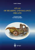
Atlas of Hearing and Balance Organs: A Practical Guide for Otolaryngologists
by Andre LeblancThe benefits of this book lie not only in the association of anatomy with modern CT and MR imaging techniques, but also and predominantly in the numerous diagrams of bony fenestration of the cochlea, the vestibule and the semicircular canals. These views reveal the membranous labyrinth, the internal organs of balance and audition, and highlight their innervation, as well as the utricular and saccular nerves of the spiral organ of corti.
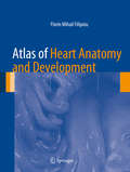
Atlas of Heart Anatomy and Development
by Florin Mihail FilipoiuThis heart anatomy book describes the cardiac development and cardiac anatomy in the development of the adult heart, and is illustrated by numerous images and examples. It contains 550 images of dissected embryo and adult hearts, obtained through the dissection and photography of 235 hearts. It has been designed to allow the rapid understanding of the key concepts and that everything should be clearly and graphically explained in one book. This is an atlas of cardiac development and anatomy of the human heart which distinguishes itself with the use of 550 images of embryonic, fetal and adult hearts and using text that is logical and concise. All the mentioned anatomical structures are shown with the use of suggestive dissection images to emphasize the details and the overall location. All the images have detailed comments, while clinical implications are suggested. The dissections of different hearts exemplify the variability of the cardiac structures. The electron and optical microscopy images are sharp and provide great fidelity. The arterial molds obtained using methyl methacrylate are illustrative and the pictures use suggestive angles. The dissections were made on human normal and pathological hearts of different ages, increasing the clinical utility of the material contained within.
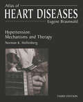
Atlas of Heart Diseases: Hypertension: Mechanisms and Therapy (Atlas of Diseases)
by Eugene Braunwald Norman HollenbergIn this edition of the Atlas of Hypertension, Dr. Norman Hollenberg and more than 20 leading authorities have worked together to capture the most updated and pertinent information in the field of hypertension. This new edition is a modern and complete visual library of up-to-date information on the most current pharmacologic and treatment advances in the field. With over 600 vivid illustrations, clinical photographs, instructive diagrams and charts, this updated reference provides a detailed and accurate insight into treatment and management of hypertension, covering a full range of topics. Together with detailed legends and extensive reference listings, the illustrations deliver comprehensive guidance to effective diagnosis and treatment of a wide breadth of these clinical challenges.
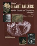
Atlas of HEART FAILURE: Cardiac Function and Dysfunction
by Wilson S. ColucciNow in its fourth edition, the Atlas of Heart Failure provides a comprehensive up-to-date overview of normal cardiac function, the mechanisms of dysfunction in heart failure, and the therapeutic approaches that are available to manage the syndrome. Designed to provide a detailed and comprehensive visual exposition of all aspects of cardiac function and dysfunction, this atlas contains several hundred images, each accompanied by detailed captions, carefully selected by expert authors, and reviewed by the editor.
Atlas of Heart Failure: Cardiac Function and Dysfunction
by Wilson S. ColucciIn its third edition, the Atlas of Heart Failure provides a comprehensive up-to-date overview of normal cardiac function, the mechanisms of dysfunction in heart failure, and the therapeutic approaches that are available to manage the syndrome. Designed to provide a detailed and comprehensive visual exposition of all aspects of cardiac function and dysfunction, this atlas contains several hundred images, each accompanied by detailed captions, carefully selected by expert authors, and reviewed by the editor.
Atlas of Hematologic Neoplasms
by Tsieh SunDue to its rapid development in recent years, hematopathology has become a very complicated discipline. The current development is mainly in two aspects: the new classification of lymphomas and leukemias and the new techniques.The Revised European-American Classification of Lymphoid Neoplasms (REAL classification) and the World Health Organization (WHO) classification of hematologic neoplasms require not only morphologic criteria but also immunophenotyping and molecular genetics for the diagnosis of hematologic tumors. Immunophenotyping is performed by either flow cytometry or immunohistochemistry. There are many new monoclonal antibodies and new equipments accumulated in recent years that make immunophenotyping more or more accurate and helpful. There are even more new techniques invented in recent years in the field of molecular genetics. In cytogenetics, the conventional karyotype is supplemented and partly replaced by the fluorescence in situ hybridization (FISH) technique. The current development of gene expression profiling is even more powerful in terms of subtyping the hematologic tumors, which may help guiding the treatment and predict the prognosis. In molecular biology, the tedious Southern blotting technique is largely replaced by polymerase chain reaction (PCR). The recent development in reverse-transcriptase PCR and quantitative PCR makes these techniques even more versatile. Because of these new developments, hematopathology has become too complicated to handle by a general pathologist. Many hospitals have to hire a newly trained hematopathologist to oversee peripheral blood, bone marrow and lymph node examinations. These young hematopathologists are geared to the new techniques, but most of them are inexperienced in morphology. No matter how well-trained a hematopathologist is, he or she still needs to see enough cases so that they can recognize the morphology and use the new techniques to substantiate the diagnosis. In other words, morphology is still the basis for the diagnosis of lymphomas and leukemias. Therefore, a good color atlas is the most helpful tool for these young hematopathologists and for the surgical pathologists who may encounter a few cases of hematologic tumors from time to time. In a busy daily practice, it is difficult to refer to a comprehensive hematologic textbook all the time. There are a few hematologic color atlases on the market to show the morphology of the normal blood cells and hematologic tumor cells. These books are helpful but not enough, because tumor cell morphology is variable from case to case and different kinds of tumor cells may look alike and need to be differentiated by other parameters. The best way to learn morphology is through the format of clinical case study. This format is also consistent with the daily practice of hematopathologists and with the pattern in all the specialty board examinations. Therefore, it is a good learning tool for the pathology residents, hematology fellows as well as medical students. This proposed book will present 83 clinical cases with clinical history, morphology of the original specimen and a list of differential diagnoses. This is followed by further testing with pictures to show the test results. At the end, a correct diagnosis is rendered with subsequent brief discussion on how the diagnosis is achieved. A few useful references will be cited and a table will be provided for differential diagnosis in some cases. The major emphasis is the provision of 500 color photos of peripheral blood smears, bone marrow aspirates, core biopsy, lymph node biopsy and biopsies of other solid organs that are involved with lymphomas and leukemias. Pictures of other diagnostic parameters, such as flow cytometric histograms, immunohistochemical stains, cytogenetic karyotypes, fluorescence in situ hybridization and polymerase chain reaction, will also be included. A comprehensive approach with consideration of clinical, morphologic, immunophenotypic and molecular genetic aspects is the
Atlas of Hepatocellular Carcinoma Pathology
by Haeryoung Kim Wei-Qiang Leow Regina Lo Paulo Giovanni Mendoza Anthony Wing-Hung ChanThis atlas is designed to be a user-friendly bench-side reference for pathology trainees and general pathologists in handling and interpreting specimens of hepatocellular carcinoma. It provides over 550 high-quality gross and microscopic photos focusing on hepatocellular carcinoma and its mimickers, and demonstrating a full range of various histological variants of hepatocellular carcinoma. Introductory text in each chapter summarises salient clinical associations, pathological features, and molecular alterations of different variants of hepatocellular carcinoma. Differentiation between hepatocellular carcinoma and its mimickers is illustrated through various case studies.The authors are nationally and internationally recognized hepatopathologists in the Asian-Pacific regions (Hong Kong, Korea, the Philippines, and Singapore), in which the incidence of hepatocellular carcinoma is high.The authors are nationally and internationally recognized hepatopathologists in the Asian-Pacific regions (Hong Kong, Korea, the Philippines, and Singapore), in which the incidence of hepatocellular carcinoma is high.The authors are nationally and internationally recognized hepatopathologists in the Asian-Pacific regions (Hong Kong, Korea, the Philippines, and Singapore), in which the incidence of hepatocellular carcinoma is high.The authors are nationally and internationally recognized hepatopathologists in the Asian-Pacific regions (Hong Kong, Korea, the Philippines, and Singapore), in which the incidence of hepatocellular carcinoma is high.The authors are nationally and internationally recognized hepatopathologists in the Asian-Pacific regions (Hong Kong, Korea, the Philippines, and Singapore), in which the incidence of hepatocellular carcinoma is high.The authors are nationally and internationally recognized hepatopathologists in the Asian-Pacific regions (Hong Kong, Korea, the Philippines, and Singapore), in which the incidence of hepatocellular carcinoma is high.The authors are nationally and internationally recognized hepatopathologists in the Asian-Pacific regions (Hong Kong, Korea, the Philippines, and Singapore), in which the incidence of hepatocellular carcinoma is high.
Atlas of High-Resolution Manometry, Impedance, and pH Monitoring
by Sarvee Moosavi Ali Rezaie Mark Pimentel Nipaporn PichetshoteThis atlas provides a concise yet comprehensive overview of high-resolution manometry, impedance and pH monitoring. Through instructive text and over 130 high-yield images, the atlas describes the basic principles of esophageal, antroduodenal and anorectal high-resolution manometry, reviews both normal and pathologic findings on manometry, covers technical aspects of pH monitoring and impedance, and outlines advances in equipment, software, and diagnostic guidelines.Written by experts in the field, Atlas of High-Resolution Manometry, Impedance, and pH Monitoring is a valuable resource for gastroenterologists and other clinicians and practitioners who work or are interested in the GI motility field.

An Atlas of Histology
by Shu-Xin ZhangBridging the gap between textbook diagrams and the complex reality of histological preparations, this magnificent atlas of human microanatomy is designed to help students understand the complex structures encountered when viewing microscopic sections of tissues. Instead of simply depicting an individual section, each drawing is a compilation of the key structures and features seen in many preparations from similar tissues or organs. Invaluable to students in a range of life science and medical disciplines including human and veterinary medicine, dentistry, mammalian biology, pharmacy, and nursing.

Atlas of Histopathology of the Cervix Uteri
by Gisela Dallenbach-Hellweg Magnus von Knebel Doeberitz Marcus J. Trunk-GemacherAn atlas covering the normal and pathologic histology of the uterine cervix. Differential diagnosis is given in detail yet related to clinical aspects, so that a functional de- scription of benefit in daily practice is achieved.

Atlas of Human Anatomy: Volumes 1-3
by F. Kiss J. SzentágothaiAFTER ten years' preparation the first edition of our Atlas of Human Anatomy was published between 1946 and 1951. Our experience enabled us to improve each of the subsequent editions and the present one has also been thoroughly revised and enlarged to allow the inclusion of more instructive illus trations. Throughout we have adhered to our original intention that this work should be a well propor tioned Atlas of life-like illustrations primarily for medical students but also useful to the practising physician and surgeon. The introduction of topographical illustrations in the third volume has been welcomed by readers and, while not embarking on histology, semi-microscopic figures have been introduced into some chapters for a better understanding of function. We did not deviate without reason from the currently accepted methods of illustrating the elements of the different systems such as bones, joints, muscles, vessels and nerves and we were at pains to base our illustrations on original dissections and to include in them only essential details. The use of colour in the illustrations, introduced by the Italian anatomist Aselli (1627), was with didactic intent. The legends to the illustrations of this edition use the nomenclature of the "Nomina Anatomica", Paris 1955 (PNA) , as revised in New York in 1960.

Atlas of Human Anatomy: Atlas of Human Anatomy: Latin Terminology E-Book (Netter Basic Science)
by Frank H. NetterThe only anatomy atlas illustrated by physicians, Atlas of Human Anatomy, 7th edition, brings you world-renowned, exquisitely clear views of the human body with a clinical perspective. In addition to the famous work of Dr. Frank Netter, you'll also find nearly 100 paintings by Dr. Carlos A. G. Machado, one of today's foremost medical illustrators. Together, these two uniquely talented physician-artists highlight the most clinically relevant views of the human body. In addition, more than 50 carefully selected radiologic images help bridge illustrated anatomy to living anatomy as seen in everyday practice. Anatomic labels follow the international standard in Latin. - Region-by-region coverage, including Muscle Table appendices at the end of each section. - Large, clear illustrations with comprehensive labels not only of major structures, but also of those with important relationships. - Tabular material in separate pages so the printed page stays focused on the illustration.Updates to the 7th Edition – based on requests from students and practitioners alike: - For the first time – a Latin-English edition. Latin nomenclature based on the international anatomic standard, Terminologia Anatomica. - New Systems Overview section featuring brand-new, full-body views of surface anatomy, vessels, nerves, and lymphatics. - More than 25 new illustrations by Dr. Machado, including the clinically important fascial columns of the neck, deep veins of the leg, hip bursae, and vasculature of the prostate; and difficult-to-visualize areas like the infratemporal fossa. - New Clinical Tables at the end of each regional section that focus on structures with high clinical significance. These tables provide quick summaries, organized by body system, and indicate where to best view key structures in the illustrated plates. - More than 50 new radiologic images – some completely new views and others using newer imaging tools – have been included based on their ability to assist readers in grasping key elements of gross anatomy. - Student Consult access includes a pincode to unlock the complete enhanced eBook of the Atlas through Student Consult.

Atlas of Human Anatomy E-Book: Digital eBook (Netter Basic Science)
by Frank H. NetterThe only anatomy atlas illustrated by physicians, Atlas of Human Anatomy, 7th edition, brings you world-renowned, exquisitely clear views of the human body with a clinical perspective. In addition to the famous work of Dr. Frank Netter, you’ll also find nearly 100 paintings by Dr. Carlos A. G. Machado, one of today’s foremost medical illustrators. Together, these two uniquely talented physician-artists highlight the most clinically relevant views of the human body. In addition, more than 50 carefully selected radiologic images help bridge illustrated anatomy to living anatomy as seen in everyday practice.Region-by-region coverage, including Muscle Table appendices at the end of each section. Large, clear illustrations with comprehensive labels not only of major structures, but also of those with important relationships.Updates to the 7th Edition – based on requests from students and practitioners alike: New Systems Overview section featuring brand-new, full-body views of surface anatomy, vessels, nerves, and lymphatics. More than 25 new illustrations by Dr. Machado, including the clinically important fascial columns of the neck, deep veins of the leg, hip bursae, and vasculature of the prostate; and difficult-to-visualize areas like the infratemporal fossa. New Clinical Tables at the end of each regional section that focus on structures with high clinical significance. These tables provide quick summaries, organized by body system, and indicate where to best view key structures in the illustrated plates. More than 50 new radiologic images – some completely new views and others using newer imaging tools – have been included based on their ability to assist readers in grasping key elements of gross anatomy. Updated terminology based on the international anatomic standard, Terminologia Anatomica, with common clinical eponyms included.
Atlas of Human Anatomy, Professional Edition E-Book: Including Student Consult Interactive Ancillaries And Guides (Netter Basic Science)
by Frank H. NetterThe gold standard of excellence for 25 years, Frank H. Netter, MD’s Atlas of Human Anatomy offers unsurpassed depictions of the human body in clear, brilliant detail – all from a clinician’s perspective. With its emphasis on anatomic relationships and clinically relevant views, Dr. Netter’s work provides a coherent, lasting visual vocabulary for understanding anatomy and how it applies to medicine today.Consult this title on your favorite e-reader. Compatible with Kindle®, nook®, and other popular devices. View anatomy from a clinical perspective with hundreds of exquisite, hand-painted illustrations created by pre-eminent medical illustrator Frank H. Netter, MD. Join the global community of medical and healthcare students and professionals who rely on Netter to optimize learning and clarify even the most difficult aspects of human anatomy. Comprehensive labeling uses the international anatomic standard terminology, Terminologia Anatomica, and every aspect of the Atlas is reviewed and overseen by clinical anatomy and anatomy education experts. Netter’s Anatomy Atlas is also available as an app for iPad®.Explore additional unique perspectives of difficult-to-visualize anatomy through all-new paintings by Dr. Carlos Machado, including breast lymph drainage; the pterygopalantine fossa; the middle ear; the path of the internal carotid artery; and the posterior knee, plus additional new plates on arteries of the limbs and new radiologic images.Master challenging structures with visual region-by-region coverage -- including Muscle Table appendices at the end of each Section.

Atlas of Human Anatomy, Professional Edition E-Book: including NetterReference.com Access with Full Downloadable Image Bank (Netter Basic Science)
by Frank H. NetterThe gold standard of excellence for 25 years, Frank H. Netter, MD’s Atlas of Human Anatomy offers unsurpassed depictions of the human body in clear, brilliant detail – all from a clinician’s perspective. With its emphasis on anatomic relationships and clinically relevant views, Dr. Netter’s work provides a coherent, lasting visual vocabulary for understanding anatomy and how it applies to medicine today. View anatomy from a clinical perspective with hundreds of exquisite, hand-painted illustrations created by pre-eminent medical illustrator Frank H. Netter, MD. Join the global community of medical and healthcare students and professionals who rely on Netter to optimize learning and clarify even the most difficult aspects of human anatomy. Comprehensive labeling uses the international anatomic standard terminology, Terminologia Anatomica, and every aspect of the Atlas is reviewed and overseen by clinical anatomy and anatomy education experts. Consulting Editors include: John T. Hansen, PhD; Brion Benninger, MD, MS; Jennifer Brueckner-Collins, PhD, Todd M. Hoagland, PhD, and R. Shane Tubbs, MS, PA-C, PhD. Explore additional unique perspectives of difficult-to-visualize anatomy through all-new paintings by Dr. Carlos Machado, including breast lymph drainage; the pterygopalantine fossa; the middle ear; the path of the internal carotid artery; and the posterior knee, plus additional new plates on arteries of the limbs and new radiologic images. Master challenging structures with visual region-by-region coverage -- including Muscle Table appendices at the end of each Section. Student Consult access includes a suite of interactive tools and guides, including selected images formatted as self-testing exercises; dissection videos; multiple choice questions; illustrated axial cross-sections and scroll-throughs; Key Point Anatomy Guides; additional plates from previous editions; and more.
Atlas of Human Body Ultrasound Scanning: Methods And Diagnostic Applications
by Mei ZhangThis atlas describes the diagnosis practice and cases of human body ultrasound. It includes anatomic section, standard scan ultrasonogram of every organ, scanning methods and key points, measurement methods, normal value ranges, and clinical significances of every sections. Providing basic information and fundamental principles of ultrasonic diagnosis, it discusses ultrasound scanning of 14 organs in individual chapters. Each section sonography is accompanied by the scanning method, section structure, measuring method and clinical application. The uniform structure and detailed instructions make this atlas an easy-to-use resource for residents to refer to when they encounter specific ultrasound diagnostic problems.

Atlas of Human Chromosome Heteromorphisms
by Herman E. Wyandt Vijay S. TonkCritical to the accurate diagnosis of human illness is the need to distinguish clinical features that fall within the normal range from those that do not. That distinction is often challenging and not infrequently requires considerable experience at the bedside. It is not surprising that accurate cytogenetic diagnosis is also often a challenge, especially when chromosome study reveals morphologic findings that raise the question of normality. Given the realization that modern human cytogenetics is just over five decades old, it is noteworthy that thorough documentation of normal chromosome var- tion has not yet been accomplished. One key diagnostic consequence of the inability to distinguish a “normal” variation in chromosome structure from a pathologic change is a missed or inaccurate diagnosis. Clinical cytogeneticists have not, however, been idle. Rather, progressive biotechnological advances coupled with virtual completion of the human genome project have yielded increasingly better microscopic resolution of chromosome structure. Witness the progress from the early short condensed chromosomes to the later visualization of chromosomes through banding techniques, hi- resolution analysis in prophase, and more recently to analysis by fluorescent in situ hybridization (FISH).
Atlas of Human Cranial Macromorphoscopic Traits
by Joseph T. Hefner Kandus C. LindeAtlas of Human Cranial Macromorphoscopic Traits synthesizes macromorphoscopic traits and their analysis in an accessible manner, providing detailed descriptions and examples of the various character state manifestations intended for use in classrooms, laboratories, and in the field. The volume begins with an outline of the macromorphoscopic dataset, its history, recent modifications to the historical approach, and recent technological and analytical advances. Additional sections cover Nomenclature, Gross Anatomy, Function, Methodology, Line Drawings, Detailed Definitions, Multiple High-resolution Photographs, and Population Variation Data from the Macromorphoscopic Databank (MaMD). The volume concludes with a chapter outlining the statistical analysis of macromorphoscopic data and a summary of the computer programs and reference databases available to forensic anthropologists for the analysis of these data.Provides detailed descriptions, illustrations and high-resolution images of various character state manifestations of seventeen macromorphoscopic traitsApplies to both forensic and bioarcheological researchWritten by the foremost expert on macromorphoscopic trait analysis and estimation of ancestry in forensic anthropology

Atlas of Human Hemopoietic Development
by E. Kelemen W. Calvo T.M. FliednerDuring the past 20 years, celJ biology has made immense strides which have completely transformed the time-honored morphological hematology of yesterday. This progress is primarily due to the introduction of new techniques which allow functional rather than anatomic studies: labeling techniques have made possible the study of celJ kinetics from birth to death of a celJ: culture techniques (both in vivo and in vitro) have made it possible to establish the progeny of certain stern celJs, their growth poten tiaL and the mechanisms of their regulation. The results have been so impressive and have so aroused the enthusiasm 01' young hematologists that it has become fashionable in so me quarters to consider the microscope an "extinct instrument" and morphology littlc more than an outmoded (if agreeable) pastime of little scientific interest. One of the consequences is the wish of some investigators to study cytology without the aid of their eyes. The present book makes us realize once more that morphology is the science of structure and shape and that its aim is not to colJect pictures but to understand them. It is true that microscopic observation, even when made with the electron microscope, cannot by itself answer some basic questions of celJ biology. However, the hematologist who uses only a single technique is like a person who would describe the world from the point of view of a single sensory organ and would refuse the aid of the others.

Atlas of Human Infectious Diseases
by Heiman F.L.Wertheim Peter Horby John P. WoodallThe Atlas of Human Infectious Diseases provides a much needed practical and visual overview of the current distribution and determinants of major infectious diseases of humans. The comprehensive full-color maps show at a glance the areas with reported infections and outbreaks, and are accompanied by a concise summary of key information on the infectious agent and its clinical and epidemiological characteristics. Since infectious diseases are dynamic, the maps are presented in the context of a changing world, and how these changes are influencing the geographical distribution on human infections. This unique atlas: Contains more than 145 high quality full-color maps covering all major human infectious diseases Provides key information on the illustrated infectious diseases Has been compiled and reviewed by an editorial board of infectious disease experts from around the world The result is a concise atlas with a consistent format throughout, where material essential for understanding the global spatial distribution of infectious diseases has been thoughtfully assembled by international experts. Atlas of Human Infectious Diseases is an essential tool for infectious disease specialists, medical microbiologists, virologists, travel medicine specialists, and public health professionals. The Atlas of Human Infectious Diseases is accompanied by a FREE enhanced Wiley Desktop Edition - an interactive digital version of the book with downloadable images and text, highlighting and note-taking facilities, book-marking, cross-referencing, in-text searching, and linking to references and glossary terms.
Atlas of Human Infectious Diseases
by Heiman F. L. Wertheim Peter Horby John P. WoodallThe Atlas of Human Infectious Diseases provides a much needed practical and visual overview of the current distribution and determinants of major infectious diseases of humans. The comprehensive full-color maps show at a glance the areas with reported infections and outbreaks, and are accompanied by a concise summary of key information on the infectious agent and its clinical and epidemiological characteristics. Since infectious diseases are dynamic, the maps are presented in the context of a changing world, and how these changes are influencing the geographical distribution on human infections. This unique atlas: Contains more than 145 high quality full-color maps covering all major human infectious diseases Provides key information on the illustrated infectious diseases Has been compiled and reviewed by an editorial board of infectious disease experts from around the world The result is a concise atlas with a consistent format throughout, where material essential for understanding the global spatial distribution of infectious diseases has been thoughtfully assembled by international experts. Atlas of Human Infectious Diseases is an essential tool for infectious disease specialists, medical microbiologists, virologists, travel medicine specialists, and public health professionals. The Atlas of Human Infectious Diseases is accompanied by a FREE enhanced Wiley Desktop Edition - an interactive digital version of the book with downloadable images and text, highlighting and note-taking facilities, book-marking, cross-referencing, in-text searching, and linking to references and glossary terms.

Atlas of Human Limb Joints
by Jacques GuyotThe aim of this book is to present as completely as possible a description of the joints of the human limbs, using photographs and drawings of anatomical dissections. Existing descriptive anatomical accounts are often theoretical and not linked to functional activity of the joints concerned; they lack practical demonstra tion of the anatomy. We hope to fill the gap in the available literature by presentation of this book. The work is directed towards anatomists, doctors interested in joint pathology, orthopaedic surgeons, rheumatologists, radiologists, specialists in sport injuries and rehabilitation, also to physicians in general, physiotherapists and students. The book is divided into seven chapters. Each chapter comprises two parts: the first is a brief account of the functional anatomy of the joint. It does not offer a complete description but an overall summary of the functional structures involved. The second section, the main part, includes illustrations (drawings and photographs of anatomical dissections). The dissections and the photographs were prepared in the Department of Anatomy directed by Professor HENRI M. DUVERNaY from cadaveric specimens preserved by the method described by WINCKLER (1964). The process of dissecting the ligamentous structures around joints has proved techni cally difficult. It is easy to create artificially structures from the mass of fibrous tissue and on a number of occasions we have been unable to locate ligaments which are described in some anatomical accounts.
Atlas of Human Limb Joints
by Jacques GuyotIn this work, the author provides the most complete description of human limb joints available today. His presentation is divided into 2 parts. The first part contains a summary of the functional anatomy of each of the joints. The second part is devoted to the pictorial illustration of the joints, consisting of photographs, drawings and diagrams of meticulously prepared dissections of the ligamentous structures surrounding the joints as well as the joints themselves. From the reviews of the first edition: "...of great importance and interest for anatomists, surgeons, specialists in sports medicine and physiotherapists, and departmental libraries must include this book. The quality of dissections, photographs and artistic diagrams must be seen to be believed. The book is higly recommended and will be of great delight to those concerned with the function and surgery of joints." The Journal of Bone and Joint Surgery#1
Atlas of Human Poisoning and Envenoming
by James H. DiazClinicians undergoing competency testing, certification, and periodic recertification are frequently faced with computer-based exams designed to evaluate clinical acumen and judgment. Test questions often include an image or radiograph followed by a vignette of the clinical encounter and a series of questions. Designed to better prepare practitione

Atlas of Human Prenatal Morphogenesis
by J.E. JirásekA little picture is worth a million words. Chinese proverb Prenatal human development is an extremely complicated process related to genetics, biochemistry, anatomy, and physiology. There are no developmental changes, either chemical or morphologic, without simultaneous changes in molecular organization. The astonishing buildup ofbiostructures always precedes their proper function. The development of an embryo is genetically coded and is based on interactions related to the selective switching on and off of genes. Interactions are cell-to-cell mediated, mediated by extracellular fluids, or mediated by special pathways. Every substance involved in developmental is to be recognized by its target. interactions, before triggering a metabolic or a morphogenic event, Complex physical and immunologic recognitions are involved in the process of differentiation. Small pieces of evidence are collected to create a mosaic picture elucidating the development. This picture is fascinating and represents the biggest biological puzzle: the puzzle of development. There is no doubt that analysis of human prenatal development is a basis for understanding normal and pathologic relationships between structure and function. Today, there are approximately 2000 different inborn congenital anomalies and syndromes. This book attempts to present a complete realistic account of human morphogenesis, the differentiation of structures, using direct photographs of normal specimens obtained from legal medical abortions of unwanted pregnancies. Emphasis has been placed on contemporary techniques: histochemistry and scanning electron microscopy. The text is as simple as possible; meticulous detailed anatomic descriptions have been omitted.